

6.2 Classification and Biogenesis of EVs
EVs are lipid bilayer-limited nano-sized vesicles, ranging from 30 to 1000 nm in
diameter [4, 5]. Although diverse nomenclatures represent for EVs derived from
three domains of life (archaea, bacteria, and eukarya) as reported in EVpedia at
http://evpedia.info (Fig. 6.3), mammalian EVs are generally categorized into
exosomes and ectosomes based on their biogenesis mechanisms: exosomes
(30–100 nm in diameter) that are released to the extracellular space after fusion of
multivesicular bodies with the plasma membrane, and ectosomes (100–1000 nm in
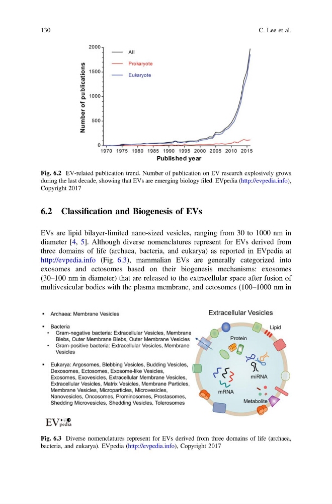
Fig. 6.2 EV-related publication trend. Number of publication on EV research explosively grows
during the last decade, showing that EVs are emerging biology filed. EVpedia (http://evpedia.info),
Copyright 2017
Fig. 6.3 Diverse nomenclatures represent for EVs derived from three domains of life (archaea,
bacteria, and eukarya). EVpedia (http://evpedia.info), Copyright 2017
130
C. Lee et al.
EVs are lipid bilayer-limited nano-sized vesicles, ranging from 30 to 1000 nm in
diameter [4, 5]. Although diverse nomenclatures represent for EVs derived from
three domains of life (archaea, bacteria, and eukarya) as reported in EVpedia at
http://evpedia.info (Fig. 6.3), mammalian EVs are generally categorized into
exosomes and ectosomes based on their biogenesis mechanisms: exosomes
(30–100 nm in diameter) that are released to the extracellular space after fusion of
multivesicular bodies with the plasma membrane, and ectosomes (100–1000 nm in
Fig. 6.2 EV-related publication trend. Number of publication on EV research explosively grows
during the last decade, showing that EVs are emerging biology filed. EVpedia (http://evpedia.info),
Copyright 2017
Fig. 6.3 Diverse nomenclatures represent for EVs derived from three domains of life (archaea,
bacteria, and eukarya). EVpedia (http://evpedia.info), Copyright 2017
130
C. Lee et al.