

diameter), also known as microvesicles, that are secreted via shedding of plasma
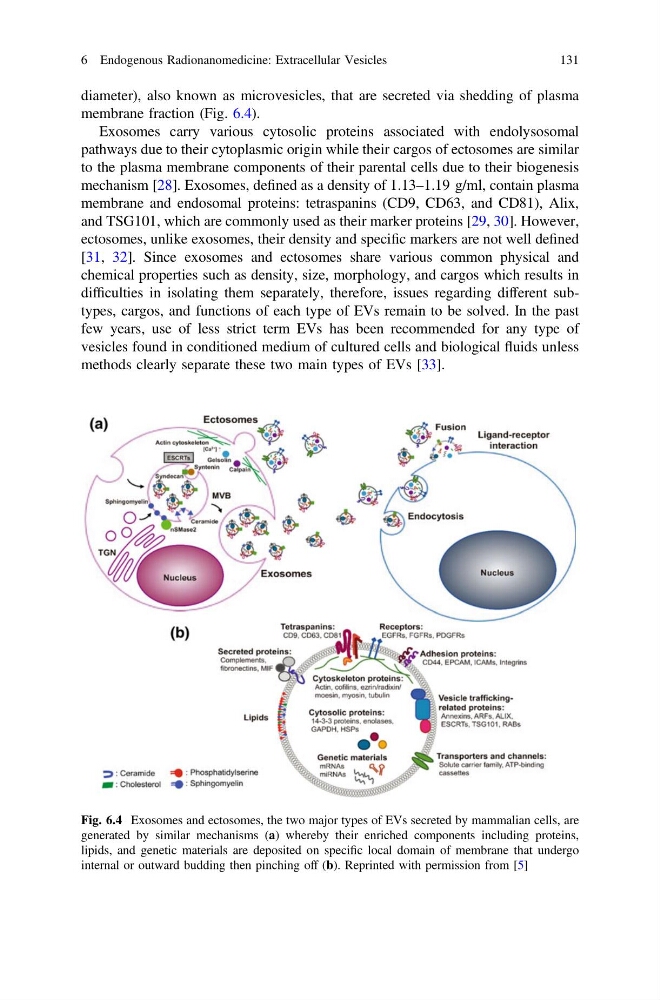
membrane fraction (Fig. 6.4).
Exosomes carry various cytosolic proteins associated with endolysosomal
pathways due to their cytoplasmic origin while their cargos of ectosomes are similar
to the plasma membrane components of their parental cells due to their biogenesis
mechanism [28]. Exosomes, defined as a density of 1.13–1.19 g/ml, contain plasma
membrane and endosomal proteins: tetraspanins (CD9, CD63, and CD81), Alix,
and TSG101, which are commonly used as their marker proteins [29, 30]. However,
ectosomes, unlike exosomes, their density and specific markers are not well defined
[31, 32]. Since exosomes and ectosomes share various common physical and
chemical properties such as density, size, morphology, and cargos which results in
difficulties in isolating them separately, therefore, issues regarding different subtypes, cargos, and functions of each type of EVs remain to be solved. In the past
few years, use of less strict term EVs has been recommended for any type of
vesicles found in conditioned medium of cultured cells and biological fluids unless
methods clearly separate these two main types of EVs [33].
Fig. 6.4 Exosomes and ectosomes, the two major types of EVs secreted by mammalian cells, are
generated by similar mechanisms (a) whereby their enriched components including proteins,
lipids, and genetic materials are deposited on specific local domain of membrane that undergo
internal or outward budding then pinching off (b). Reprinted with permission from [5]
6 Endogenous Radionanomedicine: Extracellular Vesicles
131
membrane fraction (Fig. 6.4).
Exosomes carry various cytosolic proteins associated with endolysosomal
pathways due to their cytoplasmic origin while their cargos of ectosomes are similar
to the plasma membrane components of their parental cells due to their biogenesis
mechanism [28]. Exosomes, defined as a density of 1.13–1.19 g/ml, contain plasma
membrane and endosomal proteins: tetraspanins (CD9, CD63, and CD81), Alix,
and TSG101, which are commonly used as their marker proteins [29, 30]. However,
ectosomes, unlike exosomes, their density and specific markers are not well defined
[31, 32]. Since exosomes and ectosomes share various common physical and
chemical properties such as density, size, morphology, and cargos which results in
difficulties in isolating them separately, therefore, issues regarding different subtypes, cargos, and functions of each type of EVs remain to be solved. In the past
few years, use of less strict term EVs has been recommended for any type of
vesicles found in conditioned medium of cultured cells and biological fluids unless
methods clearly separate these two main types of EVs [33].
Fig. 6.4 Exosomes and ectosomes, the two major types of EVs secreted by mammalian cells, are
generated by similar mechanisms (a) whereby their enriched components including proteins,
lipids, and genetic materials are deposited on specific local domain of membrane that undergo
internal or outward budding then pinching off (b). Reprinted with permission from [5]
6 Endogenous Radionanomedicine: Extracellular Vesicles
131