

can guide minimal invasive surgery in cancers with early stage such as breast
cancer, melanoma, and etc.
99m Tc nanocolloidal albumin was approved in Europe
for lymphoscintigraphy, bone marrow scintigraphy and visualization of inflammatory processes. Manufacturers declare in the technical leaflet that more than 95%
of the particles of nanocolloidal albumin are smaller than 80 nm. Three commercially available
99m Tc HSA nanocolloids (Nanoalbumon
® , Nanocoll
® and
Nanotop
® ) were compared for their size and radioactivity distribution [34]. The
ranges of particle diameter were 6.5–68 nm for Nanocoll
® , 5.6–79 nm for
Nanotop
® , and 12–122 nm for Nanoalbumon
® . The size and radioactivity distribution are similar between Nanotop
® and Nanocoll
® , and those of Nanoalbumon
®
is bigger than the others. Smallest particles can pass SLNs, and more node tiers are
visualized, decreasing the accuracy of SLN mapping.
Combination of chemotherapy and radionuclide therapy based on albumin NPs
was tried to show synergistic treatment effect in a mouse model bearing murine
breast cancer 4T1 tumor [35]. HSA was labeled with radionuclide
131 I through a
standard chloramine-T oxidation method, and then were added with PTX to prepare
131 I-HSA-PTX NPs.
131 I-HSA-PTX showed obvious tumor accumulation on
gamma imaging 24 h after intravenous injection. Compared to radionuclide therapy
(
131 I-HSA) or chemotherapy (HSA-PTX) alone group, the combination therapy by
131 I-HSA-PTX was significantly more effective to decrease the tumor volumes.
Strategies labeling innate albumin in vivo were also tried using albumin binding
molecules [36]. Evans Blue (EB), an albumin binding dye, displays reversible
binding to serum albumin with IC 50 in the micromolar range. A truncated EB was
labeled with
68 Ga through 1,4,7-triazacyclononane-N,N′,N″-triacetic acid (NOTA)
chelator (
68 Ga-NEB).
68 Ga-NEB was retained in the blood circulation and cleared
slowly but steadily from the blood in healthy volunteers. As a blood volume
imaging agent,
68 Ga-NEB imaging clearly differentiated hepatic hemangioma from
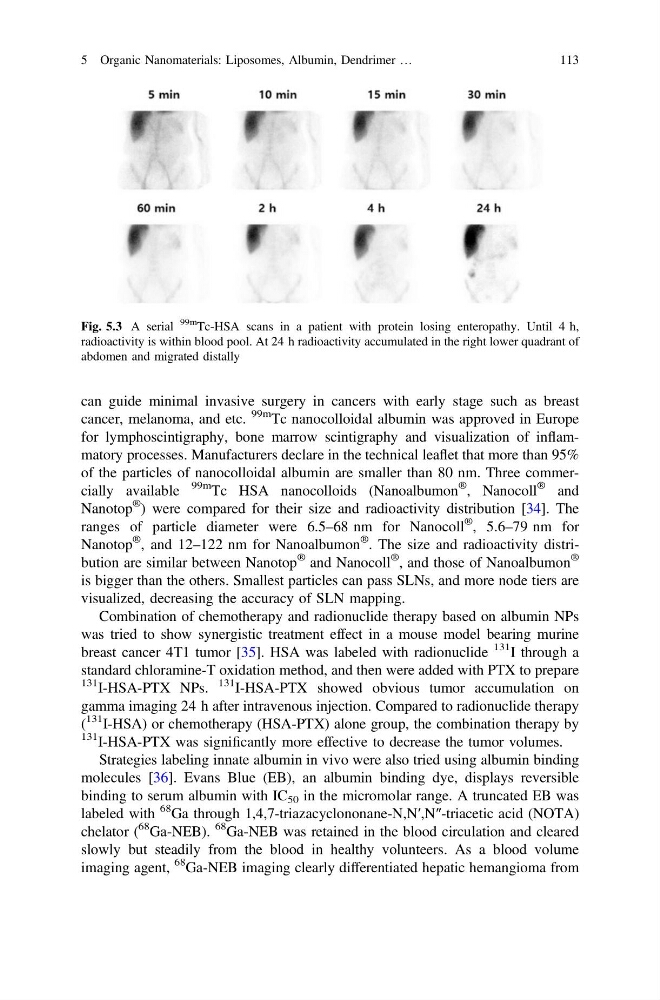
Fig. 5.3 A serial
99m
Tc-HSA scans in a patient with protein losing enteropathy. Until 4 h,
radioactivity is within blood pool. At 24 h radioactivity accumulated in the right lower quadrant of
abdomen and migrated distally
5 Organic Nanomaterials: Liposomes, Albumin, Dendrimer …
113
cancer, melanoma, and etc.
99m Tc nanocolloidal albumin was approved in Europe
for lymphoscintigraphy, bone marrow scintigraphy and visualization of inflammatory processes. Manufacturers declare in the technical leaflet that more than 95%
of the particles of nanocolloidal albumin are smaller than 80 nm. Three commercially available
99m Tc HSA nanocolloids (Nanoalbumon
® , Nanocoll
® and
Nanotop
® ) were compared for their size and radioactivity distribution [34]. The
ranges of particle diameter were 6.5–68 nm for Nanocoll
® , 5.6–79 nm for
Nanotop
® , and 12–122 nm for Nanoalbumon
® . The size and radioactivity distribution are similar between Nanotop
® and Nanocoll
® , and those of Nanoalbumon
®
is bigger than the others. Smallest particles can pass SLNs, and more node tiers are
visualized, decreasing the accuracy of SLN mapping.
Combination of chemotherapy and radionuclide therapy based on albumin NPs
was tried to show synergistic treatment effect in a mouse model bearing murine
breast cancer 4T1 tumor [35]. HSA was labeled with radionuclide
131 I through a
standard chloramine-T oxidation method, and then were added with PTX to prepare
131 I-HSA-PTX NPs.
131 I-HSA-PTX showed obvious tumor accumulation on
gamma imaging 24 h after intravenous injection. Compared to radionuclide therapy
(
131 I-HSA) or chemotherapy (HSA-PTX) alone group, the combination therapy by
131 I-HSA-PTX was significantly more effective to decrease the tumor volumes.
Strategies labeling innate albumin in vivo were also tried using albumin binding
molecules [36]. Evans Blue (EB), an albumin binding dye, displays reversible
binding to serum albumin with IC 50 in the micromolar range. A truncated EB was
labeled with
68 Ga through 1,4,7-triazacyclononane-N,N′,N″-triacetic acid (NOTA)
chelator (
68 Ga-NEB).
68 Ga-NEB was retained in the blood circulation and cleared
slowly but steadily from the blood in healthy volunteers. As a blood volume
imaging agent,
68 Ga-NEB imaging clearly differentiated hepatic hemangioma from
Fig. 5.3 A serial
99m
Tc-HSA scans in a patient with protein losing enteropathy. Until 4 h,
radioactivity is within blood pool. At 24 h radioactivity accumulated in the right lower quadrant of
abdomen and migrated distally
5 Organic Nanomaterials: Liposomes, Albumin, Dendrimer …
113