

sample has been applied to a wetted carbon film on an EM grid
(see Note 24).
2. A 1.7 μL droplet of sample buffer with 4% trehalose is applied
onto Parafilm (see Note 25).
3. The grid is rotated by 180
and firmly placed on a single layer of
Millipore DAWP membrane filter paper (0.65 μm pore size)
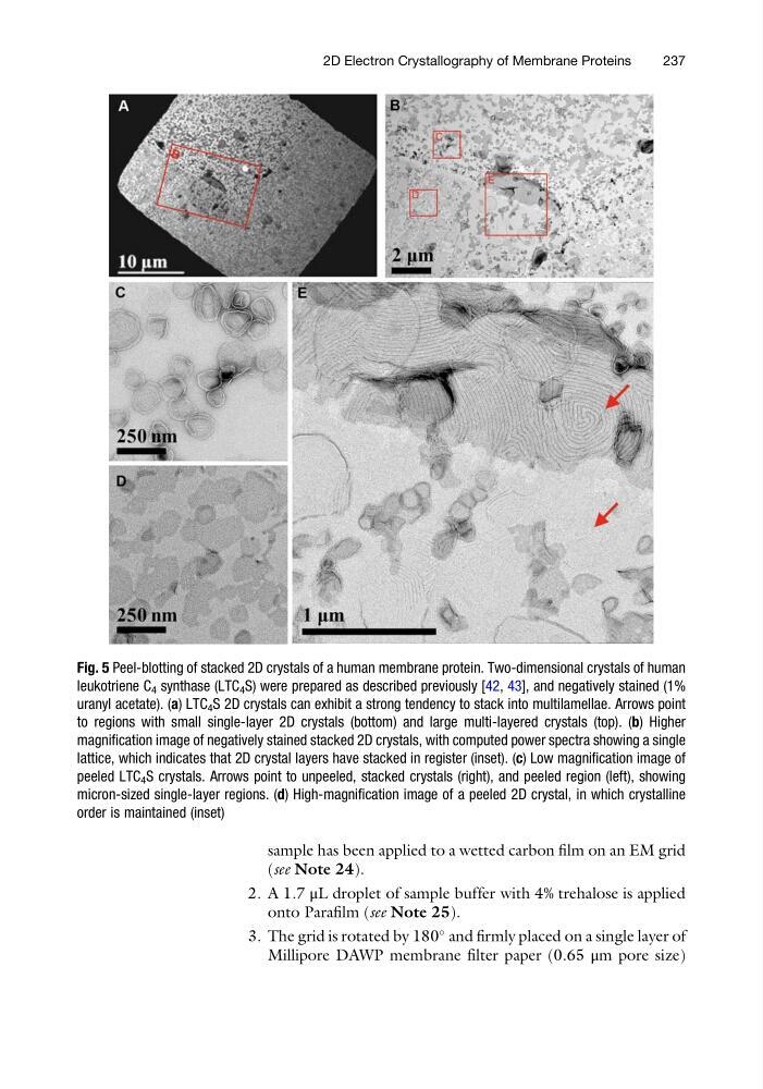
Fig. 5 Peel-blotting of stacked 2D crystals of a human membrane protein. Two-dimensional crystals of human
leukotriene C 4 synthase (LTC 4 S) were prepared as described previously [42, 43], and negatively stained (1%
uranyl acetate). (a) LTC 4 S 2D crystals can exhibit a strong tendency to stack into multilamellae. Arrows point
to regions with small single-layer 2D crystals (bottom) and large multi-layered crystals (top). (b) Higher
magnification image of negatively stained stacked 2D crystals, with computed power spectra showing a single
lattice, which indicates that 2D crystal layers have stacked in register (inset). (c) Low magnification image of
peeled LTC 4 S crystals. Arrows point to unpeeled, stacked crystals (right), and peeled region (left), showing
micron-sized single-layer regions. (d) High-magnification image of a peeled 2D crystal, in which crystalline
order is maintained (inset)
2D Electron Crystallography of Membrane Proteins
237
(see Note 24).
2. A 1.7 μL droplet of sample buffer with 4% trehalose is applied
onto Parafilm (see Note 25).
3. The grid is rotated by 180
and firmly placed on a single layer of
Millipore DAWP membrane filter paper (0.65 μm pore size)
Fig. 5 Peel-blotting of stacked 2D crystals of a human membrane protein. Two-dimensional crystals of human
leukotriene C 4 synthase (LTC 4 S) were prepared as described previously [42, 43], and negatively stained (1%
uranyl acetate). (a) LTC 4 S 2D crystals can exhibit a strong tendency to stack into multilamellae. Arrows point
to regions with small single-layer 2D crystals (bottom) and large multi-layered crystals (top). (b) Higher
magnification image of negatively stained stacked 2D crystals, with computed power spectra showing a single
lattice, which indicates that 2D crystal layers have stacked in register (inset). (c) Low magnification image of
peeled LTC 4 S crystals. Arrows point to unpeeled, stacked crystals (right), and peeled region (left), showing
micron-sized single-layer regions. (d) High-magnification image of a peeled 2D crystal, in which crystalline
order is maintained (inset)
2D Electron Crystallography of Membrane Proteins
237