

1. Following positive identification of multilamellae by negative
stain, carry out the cryo-EM grid preparation by back-injection
according to the previous section, up to step 12, in which
I.
II.
III.
IV.
V.
VI.
Float carbon film
on buffer, pick up
with grid, add
sample
Sample adheres
to carbon film
Firmly adhere
carbon film and
grid surface
Briefly dry, plunge
into cryogen
Gently separate
carbon film and
grid surface
Blot, carbon film
flattens, settles
on grid
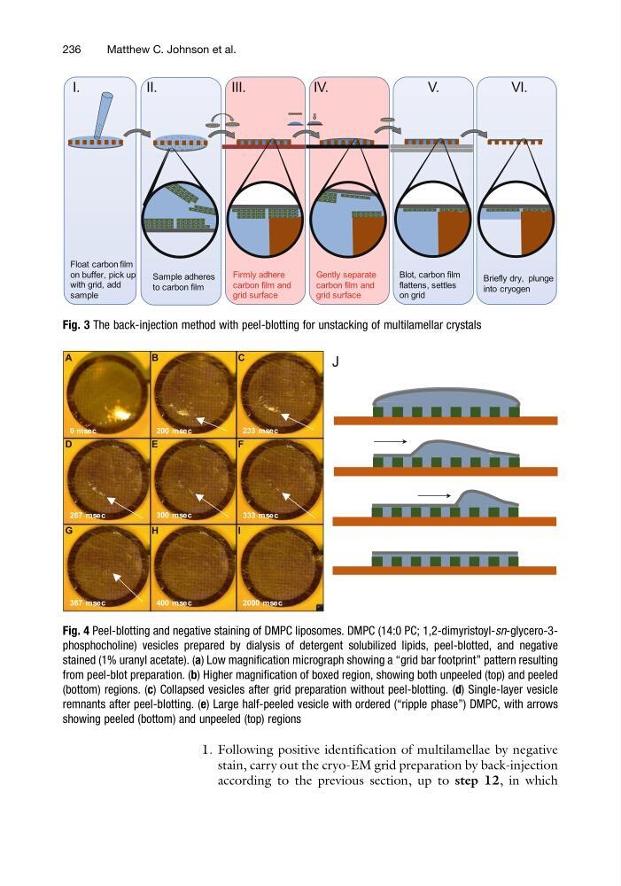
Fig. 3 The back-injection method with peel-blotting for unstacking of multilamellar crystals
A
B
C
D
E
F
G
H
I
0 msec
200 msec
233 msec
267 msec
300 msec
333 msec
367 msec
400 msec
2000 msec
J
Fig. 4 Peel-blotting and negative staining of DMPC liposomes. DMPC (14:0 PC; 1,2-dimyristoyl-sn-glycero-3phosphocholine) vesicles prepared by dialysis of detergent solubilized lipids, peel-blotted, and negative
stained (1% uranyl acetate). (a) Low magnification micrograph showing a “grid bar footprint” pattern resulting
from peel-blot preparation. (b) Higher magnification of boxed region, showing both unpeeled (top) and peeled
(bottom) regions. (c) Collapsed vesicles after grid preparation without peel-blotting. (d) Single-layer vesicle
remnants after peel-blotting. (e) Large half-peeled vesicle with ordered (“ripple phase”) DMPC, with arrows
showing peeled (bottom) and unpeeled (top) regions
236
Matthew C. Johnson et al.
stain, carry out the cryo-EM grid preparation by back-injection
according to the previous section, up to step 12, in which
I.
II.
III.
IV.
V.
VI.
Float carbon film
on buffer, pick up
with grid, add
sample
Sample adheres
to carbon film
Firmly adhere
carbon film and
grid surface
Briefly dry, plunge
into cryogen
Gently separate
carbon film and
grid surface
Blot, carbon film
flattens, settles
on grid
Fig. 3 The back-injection method with peel-blotting for unstacking of multilamellar crystals
A
B
C
D
E
F
G
H
I
0 msec
200 msec
233 msec
267 msec
300 msec
333 msec
367 msec
400 msec
2000 msec
J
Fig. 4 Peel-blotting and negative staining of DMPC liposomes. DMPC (14:0 PC; 1,2-dimyristoyl-sn-glycero-3phosphocholine) vesicles prepared by dialysis of detergent solubilized lipids, peel-blotted, and negative
stained (1% uranyl acetate). (a) Low magnification micrograph showing a “grid bar footprint” pattern resulting
from peel-blot preparation. (b) Higher magnification of boxed region, showing both unpeeled (top) and peeled
(bottom) regions. (c) Collapsed vesicles after grid preparation without peel-blotting. (d) Single-layer vesicle
remnants after peel-blotting. (e) Large half-peeled vesicle with ordered (“ripple phase”) DMPC, with arrows
showing peeled (bottom) and unpeeled (top) regions
236
Matthew C. Johnson et al.