

3.4.2 RIBFIND
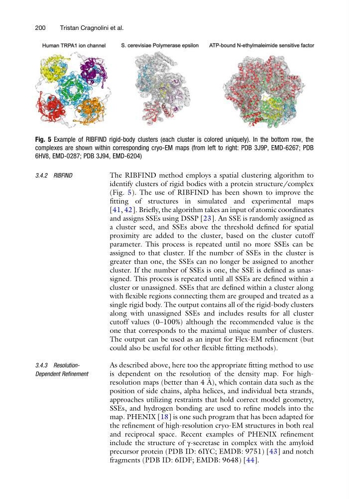
The RIBFIND method employs a spatial clustering algorithm to
identify clusters of rigid bodies with a protein structure/complex
(Fig. 5). The use of RIBFIND has been shown to improve the
fitting of structures in simulated and experimental maps
[41, 42]. Briefly, the algorithm takes an input of atomic coordinates
and assigns SSEs using DSSP [23]. An SSE is randomly assigned as
a cluster seed, and SSEs above the threshold defined for spatial
proximity are added to the cluster, based on the cluster cutoff
parameter. This process is repeated until no more SSEs can be
assigned to that cluster. If the number of SSEs in the cluster is
greater than one, the SSEs can no longer be assigned to another
cluster. If the number of SSEs is one, the SSE is defined as unassigned. This process is repeated until all SSEs are defined within a
cluster or unassigned. SSEs that are defined within a cluster along
with flexible regions connecting them are grouped and treated as a
single rigid body. The output contains all of the rigid-body clusters
along with unassigned SSEs and includes results for all cluster
cutoff values (0–100%) although the recommended value is the
one that corresponds to the maximal unique number of clusters.
The output can be used as an input for Flex-EM refinement (but
could also be useful for other flexible fitting methods).
3.4.3 ResolutionDependent Refinement
As described above, here too the appropriate fitting method to use
is dependent on the resolution of the density map. For highresolution maps (better than 4 A ˚ ), which contain data such as the
position of side chains, alpha helices, and individual beta strands,
approaches utilizing restraints that hold correct model geometry,
SSEs, and hydrogen bonding are used to refine models into the
map. PHENIX [18] is one such program that has been adapted for
the refinement of high-resolution cryo-EM structures in both real
and reciprocal space. Recent examples of PHENIX refinement
include the structure of γ-secretase in complex with the amyloid
precursor protein (PDB ID: 6IYC; EMDB: 9751) [43] and notch
fragments (PDB ID: 6IDF; EMDB: 9648) [44].
Fig. 5 Example of RIBFIND rigid-body clusters (each cluster is colored uniquely). In the bottom row, the
complexes are shown within corresponding cryo-EM maps (from left to right: PDB 3J9P, EMD-6267; PDB
6HV8, EMD-0287; PDB 3J94, EMD-6204)
200
Tristan Cragnolini et al.
The RIBFIND method employs a spatial clustering algorithm to
identify clusters of rigid bodies with a protein structure/complex
(Fig. 5). The use of RIBFIND has been shown to improve the
fitting of structures in simulated and experimental maps
[41, 42]. Briefly, the algorithm takes an input of atomic coordinates
and assigns SSEs using DSSP [23]. An SSE is randomly assigned as
a cluster seed, and SSEs above the threshold defined for spatial
proximity are added to the cluster, based on the cluster cutoff
parameter. This process is repeated until no more SSEs can be
assigned to that cluster. If the number of SSEs in the cluster is
greater than one, the SSEs can no longer be assigned to another
cluster. If the number of SSEs is one, the SSE is defined as unassigned. This process is repeated until all SSEs are defined within a
cluster or unassigned. SSEs that are defined within a cluster along
with flexible regions connecting them are grouped and treated as a
single rigid body. The output contains all of the rigid-body clusters
along with unassigned SSEs and includes results for all cluster
cutoff values (0–100%) although the recommended value is the
one that corresponds to the maximal unique number of clusters.
The output can be used as an input for Flex-EM refinement (but
could also be useful for other flexible fitting methods).
3.4.3 ResolutionDependent Refinement
As described above, here too the appropriate fitting method to use
is dependent on the resolution of the density map. For highresolution maps (better than 4 A ˚ ), which contain data such as the
position of side chains, alpha helices, and individual beta strands,
approaches utilizing restraints that hold correct model geometry,
SSEs, and hydrogen bonding are used to refine models into the
map. PHENIX [18] is one such program that has been adapted for
the refinement of high-resolution cryo-EM structures in both real
and reciprocal space. Recent examples of PHENIX refinement
include the structure of γ-secretase in complex with the amyloid
precursor protein (PDB ID: 6IYC; EMDB: 9751) [43] and notch
fragments (PDB ID: 6IDF; EMDB: 9648) [44].
Fig. 5 Example of RIBFIND rigid-body clusters (each cluster is colored uniquely). In the bottom row, the
complexes are shown within corresponding cryo-EM maps (from left to right: PDB 3J9P, EMD-6267; PDB
6HV8, EMD-0287; PDB 3J94, EMD-6204)
200
Tristan Cragnolini et al.