

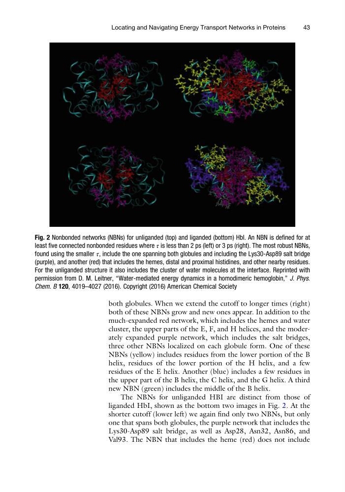
both globules. When we extend the cutoff to longer times (right)
both of these NBNs grow and new ones appear. In addition to the
much-expanded red network, which includes the hemes and water
cluster, the upper parts of the E, F, and H helices, and the moderately expanded purple network, which includes the salt bridges,
three other NBNs localized on each globule form. One of these
NBNs (yellow) includes residues from the lower portion of the B
helix, residues of the lower portion of the H helix, and a few
residues of the E helix. Another (blue) includes a few residues in
the upper part of the B helix, the C helix, and the G helix. A third
new NBN (green) includes the middle of the B helix.
The NBNs for unliganded HBI are distinct from those of
liganded HbI, shown as the bottom two images in Fig. 2. At the
shorter cutoff (lower left) we again find only two NBNs, but only
one that spans both globules, the purple network that includes the
Lys30-Asp89 salt bridge, as well as Asp28, Asn32, Asn86, and
Val93. The NBN that includes the heme (red) does not include
Fig. 2 Nonbonded networks (NBNs) for unliganded (top) and liganded (bottom) HbI. An NBN is defined for at
least five connected nonbonded residues where τ is less than 2 ps (left) or 3 ps (right). The most robust NBNs,
found using the smaller τ, include the one spanning both globules and including the Lys30-Asp89 salt bridge
(purple), and another (red) that includes the hemes, distal and proximal histidines, and other nearby residues.
For the unliganded structure it also includes the cluster of water molecules at the interface. Reprinted with
permission from D. M. Leitner, “Water-mediated energy dynamics in a homodimeric hemoglobin,” J. Phys.
Chem. B 120, 4019–4027 (2016). Copyright (2016) American Chemical Society
Locating and Navigating Energy Transport Networks in Proteins
43
both of these NBNs grow and new ones appear. In addition to the
much-expanded red network, which includes the hemes and water
cluster, the upper parts of the E, F, and H helices, and the moderately expanded purple network, which includes the salt bridges,
three other NBNs localized on each globule form. One of these
NBNs (yellow) includes residues from the lower portion of the B
helix, residues of the lower portion of the H helix, and a few
residues of the E helix. Another (blue) includes a few residues in
the upper part of the B helix, the C helix, and the G helix. A third
new NBN (green) includes the middle of the B helix.
The NBNs for unliganded HBI are distinct from those of
liganded HbI, shown as the bottom two images in Fig. 2. At the
shorter cutoff (lower left) we again find only two NBNs, but only
one that spans both globules, the purple network that includes the
Lys30-Asp89 salt bridge, as well as Asp28, Asn32, Asn86, and
Val93. The NBN that includes the heme (red) does not include
Fig. 2 Nonbonded networks (NBNs) for unliganded (top) and liganded (bottom) HbI. An NBN is defined for at
least five connected nonbonded residues where τ is less than 2 ps (left) or 3 ps (right). The most robust NBNs,
found using the smaller τ, include the one spanning both globules and including the Lys30-Asp89 salt bridge
(purple), and another (red) that includes the hemes, distal and proximal histidines, and other nearby residues.
For the unliganded structure it also includes the cluster of water molecules at the interface. Reprinted with
permission from D. M. Leitner, “Water-mediated energy dynamics in a homodimeric hemoglobin,” J. Phys.
Chem. B 120, 4019–4027 (2016). Copyright (2016) American Chemical Society
Locating and Navigating Energy Transport Networks in Proteins
43