

Hinge 1
Folate
GH Loop
GH Loop
Hinge 2
Met20 Loop
NADP+
a
b
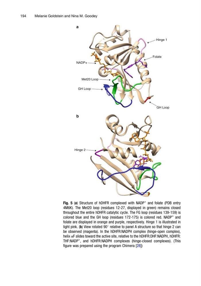
Fig. 5 (a) Structure of hDHFR complexed with NADP
+
and folate (PDB entry
4M6K). The Met20 loop (residues 12-27, displayed in green) remains closed
throughout the entire hDHFR catalytic cycle. The FG loop (residues 139-159) is
colored blue and the GH loop (residues 172-175) is colored red. NADP
+
and
folate are displayed in orange and purple, respectively. Hinge 1 is illustrated in
light pink. (b) View rotated 90
relative to panel A structure so that hinge 2 can
be observed (magenta). In the hDHFR:NADPH complex (hinge-open complex),
helix αF slides toward the active site, relative to the hDHFR:DHF:NADPH, hDHFR:
THF:NADP
+
, and hDHFR:NADPH complexes (hinge-closed complexes). (This
figure was prepared using the program Chimera [28])
194
Melanie Goldstein and Nina M. Goodey
Folate
GH Loop
GH Loop
Hinge 2
Met20 Loop
NADP+
a
b
Fig. 5 (a) Structure of hDHFR complexed with NADP
+
and folate (PDB entry
4M6K). The Met20 loop (residues 12-27, displayed in green) remains closed
throughout the entire hDHFR catalytic cycle. The FG loop (residues 139-159) is
colored blue and the GH loop (residues 172-175) is colored red. NADP
+
and
folate are displayed in orange and purple, respectively. Hinge 1 is illustrated in
light pink. (b) View rotated 90
relative to panel A structure so that hinge 2 can
be observed (magenta). In the hDHFR:NADPH complex (hinge-open complex),
helix αF slides toward the active site, relative to the hDHFR:DHF:NADPH, hDHFR:
THF:NADP
+
, and hDHFR:NADPH complexes (hinge-closed complexes). (This
figure was prepared using the program Chimera [28])
194
Melanie Goldstein and Nina M. Goodey