

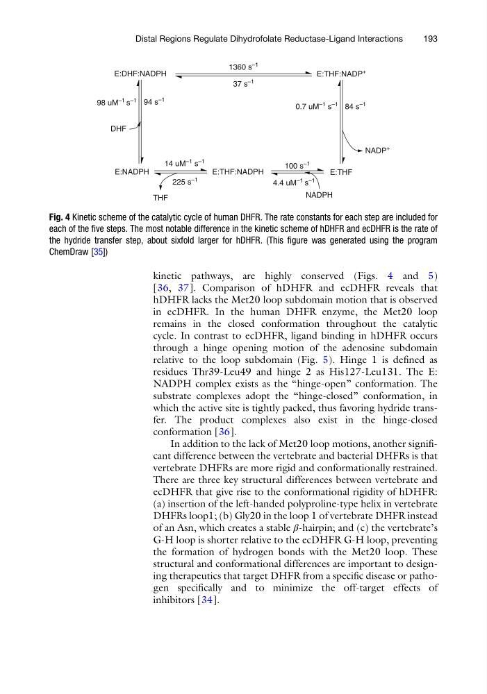
kinetic pathways, are highly conserved (Figs. 4 and 5)
[36, 37]. Comparison of hDHFR and ecDHFR reveals that
hDHFR lacks the Met20 loop subdomain motion that is observed
in ecDHFR. In the human DHFR enzyme, the Met20 loop
remains in the closed conformation throughout the catalytic
cycle. In contrast to ecDHFR, ligand binding in hDHFR occurs
through a hinge opening motion of the adenosine subdomain
relative to the loop subdomain (Fig. 5). Hinge 1 is defined as
residues Thr39-Leu49 and hinge 2 as His127-Leu131. The E:
NADPH complex exists as the “hinge-open” conformation. The
substrate complexes adopt the “hinge-closed” conformation, in
which the active site is tightly packed, thus favoring hydride transfer. The product complexes also exist in the hinge-closed
conformation [36].
In addition to the lack of Met20 loop motions, another significant difference between the vertebrate and bacterial DHFRs is that
vertebrate DHFRs are more rigid and conformationally restrained.
There are three key structural differences between vertebrate and
ecDHFR that give rise to the conformational rigidity of hDHFR:
(a) insertion of the left-handed polyproline-type helix in vertebrate
DHFRs loop1; (b) Gly20 in the loop 1 of vertebrate DHFR instead
of an Asn, which creates a stable β-hairpin; and (c) the vertebrate’s
G-H loop is shorter relative to the ecDHFR G-H loop, preventing
the formation of hydrogen bonds with the Met20 loop. These
structural and conformational differences are important to designing therapeutics that target DHFR from a specific disease or pathogen specifically and to minimize the off-target effects of
inhibitors [34].
THF
E:NADPH
DHF
98 uM
–1 s
–1 94 s
–1
E:DHF:NADPH
1360 s
–1
37 s
–1
E:THF:NADP
+
84 s
–1
0.7 uM
–1 s
–1
NADP
+
E:THF
225 s
–1
14 uM
–1 s
–1
E:THF:NADPH
4.4 uM
–1 s
–1
100 s
–1
NADPH
Fig. 4 Kinetic scheme of the catalytic cycle of human DHFR. The rate constants for each step are included for
each of the five steps. The most notable difference in the kinetic scheme of hDHFR and ecDHFR is the rate of
the hydride transfer step, about sixfold larger for hDHFR. (This figure was generated using the program
ChemDraw [35])
Distal Regions Regulate Dihydrofolate Reductase-Ligand Interactions
193
[36, 37]. Comparison of hDHFR and ecDHFR reveals that
hDHFR lacks the Met20 loop subdomain motion that is observed
in ecDHFR. In the human DHFR enzyme, the Met20 loop
remains in the closed conformation throughout the catalytic
cycle. In contrast to ecDHFR, ligand binding in hDHFR occurs
through a hinge opening motion of the adenosine subdomain
relative to the loop subdomain (Fig. 5). Hinge 1 is defined as
residues Thr39-Leu49 and hinge 2 as His127-Leu131. The E:
NADPH complex exists as the “hinge-open” conformation. The
substrate complexes adopt the “hinge-closed” conformation, in
which the active site is tightly packed, thus favoring hydride transfer. The product complexes also exist in the hinge-closed
conformation [36].
In addition to the lack of Met20 loop motions, another significant difference between the vertebrate and bacterial DHFRs is that
vertebrate DHFRs are more rigid and conformationally restrained.
There are three key structural differences between vertebrate and
ecDHFR that give rise to the conformational rigidity of hDHFR:
(a) insertion of the left-handed polyproline-type helix in vertebrate
DHFRs loop1; (b) Gly20 in the loop 1 of vertebrate DHFR instead
of an Asn, which creates a stable β-hairpin; and (c) the vertebrate’s
G-H loop is shorter relative to the ecDHFR G-H loop, preventing
the formation of hydrogen bonds with the Met20 loop. These
structural and conformational differences are important to designing therapeutics that target DHFR from a specific disease or pathogen specifically and to minimize the off-target effects of
inhibitors [34].
THF
E:NADPH
DHF
98 uM
–1 s
–1 94 s
–1
E:DHF:NADPH
1360 s
–1
37 s
–1
E:THF:NADP
+
84 s
–1
0.7 uM
–1 s
–1
NADP
+
E:THF
225 s
–1
14 uM
–1 s
–1
E:THF:NADPH
4.4 uM
–1 s
–1
100 s
–1
NADPH
Fig. 4 Kinetic scheme of the catalytic cycle of human DHFR. The rate constants for each step are included for
each of the five steps. The most notable difference in the kinetic scheme of hDHFR and ecDHFR is the rate of
the hydride transfer step, about sixfold larger for hDHFR. (This figure was generated using the program
ChemDraw [35])
Distal Regions Regulate Dihydrofolate Reductase-Ligand Interactions
193