

2.3.2 Plasmodial
DHFR-TS
A major difference between protozoal DHFR, such as Plasmodium
falciparum (pfDHFR) and Plasmodium vivax (pvDHFR), and the
DHFRs from other species is that the Plasmodium enzyme exists as
a bifunctional enzyme called dihydrofolate reductase-thymidylate
synthase (DHFR-TS) in which DHFR and TS are two domains of a
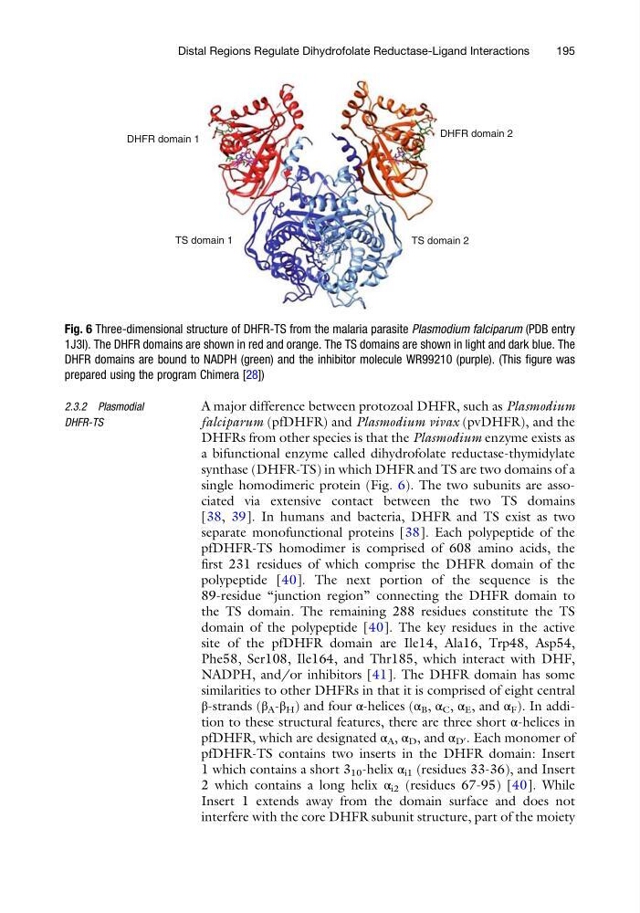
single homodimeric protein (Fig. 6). The two subunits are associated via extensive contact between the two TS domains
[38, 39]. In humans and bacteria, DHFR and TS exist as two
separate monofunctional proteins [38]. Each polypeptide of the
pfDHFR-TS homodimer is comprised of 608 amino acids, the
first 231 residues of which comprise the DHFR domain of the
polypeptide [40]. The next portion of the sequence is the
89-residue “junction region” connecting the DHFR domain to
the TS domain. The remaining 288 residues constitute the TS
domain of the polypeptide [40]. The key residues in the active
site of the pfDHFR domain are Ile14, Ala16, Trp48, Asp54,
Phe58, Ser108, Ile164, and Thr185, which interact with DHF,
NADPH, and/or inhibitors [41]. The DHFR domain has some
similarities to other DHFRs in that it is comprised of eight central
β-strands (β A -β H ) and four α-helices (α B , α C , α E , and α F ). In addition to these structural features, there are three short α-helices in
pfDHFR, which are designated α A , α D , and α D
0 . Each monomer of
pfDHFR-TS contains two inserts in the DHFR domain: Insert
1 which contains a short 3 10 -helix α i1 (residues 33-36), and Insert
2 which contains a long helix α i2 (residues 67-95) [40]. While
Insert 1 extends away from the domain surface and does not
interfere with the core DHFR subunit structure, part of the moiety
DHFR domain 1
DHFR domain 2
TS domain 1
TS domain 2
Fig. 6 Three-dimensional structure of DHFR-TS from the malaria parasite Plasmodium falciparum (PDB entry
1J3I). The DHFR domains are shown in red and orange. The TS domains are shown in light and dark blue. The
DHFR domains are bound to NADPH (green) and the inhibitor molecule WR99210 (purple). (This figure was
prepared using the program Chimera [28])
Distal Regions Regulate Dihydrofolate Reductase-Ligand Interactions
195
DHFR-TS
A major difference between protozoal DHFR, such as Plasmodium
falciparum (pfDHFR) and Plasmodium vivax (pvDHFR), and the
DHFRs from other species is that the Plasmodium enzyme exists as
a bifunctional enzyme called dihydrofolate reductase-thymidylate
synthase (DHFR-TS) in which DHFR and TS are two domains of a
single homodimeric protein (Fig. 6). The two subunits are associated via extensive contact between the two TS domains
[38, 39]. In humans and bacteria, DHFR and TS exist as two
separate monofunctional proteins [38]. Each polypeptide of the
pfDHFR-TS homodimer is comprised of 608 amino acids, the
first 231 residues of which comprise the DHFR domain of the
polypeptide [40]. The next portion of the sequence is the
89-residue “junction region” connecting the DHFR domain to
the TS domain. The remaining 288 residues constitute the TS
domain of the polypeptide [40]. The key residues in the active
site of the pfDHFR domain are Ile14, Ala16, Trp48, Asp54,
Phe58, Ser108, Ile164, and Thr185, which interact with DHF,
NADPH, and/or inhibitors [41]. The DHFR domain has some
similarities to other DHFRs in that it is comprised of eight central
β-strands (β A -β H ) and four α-helices (α B , α C , α E , and α F ). In addition to these structural features, there are three short α-helices in
pfDHFR, which are designated α A , α D , and α D
0 . Each monomer of
pfDHFR-TS contains two inserts in the DHFR domain: Insert
1 which contains a short 3 10 -helix α i1 (residues 33-36), and Insert
2 which contains a long helix α i2 (residues 67-95) [40]. While
Insert 1 extends away from the domain surface and does not
interfere with the core DHFR subunit structure, part of the moiety
DHFR domain 1
DHFR domain 2
TS domain 1
TS domain 2
Fig. 6 Three-dimensional structure of DHFR-TS from the malaria parasite Plasmodium falciparum (PDB entry
1J3I). The DHFR domains are shown in red and orange. The TS domains are shown in light and dark blue. The
DHFR domains are bound to NADPH (green) and the inhibitor molecule WR99210 (purple). (This figure was
prepared using the program Chimera [28])
Distal Regions Regulate Dihydrofolate Reductase-Ligand Interactions
195