

scale. In experimental conditions, values for FRET efficiencies are
typically between $50% and 5%, thus R 0 R 1
1
2 R 0 since FRET
pairs are around 5 nm for R 0 .
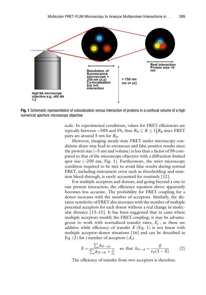
However, imaging steady-state FRET under microscopy conditions alone may lead to erroneous and false-positive results since
the protein size (~5 nm and volume) is less than a factor of 50 compared to that of the microscope objective with a diffraction-limited
spot size (~250 nm, Fig. 1). Furthermore, the strict microscopy
condition required to be met to avoid false results during normal
FRET, including instrument error such as thresholding and emission bleed-through, is rarely accounted for routinely [12].
For multiple acceptors and donors, and going beyond a one to
one protein interaction, the efficiency equation above apparently
becomes less accurate. The probability for FRET coupling for a
donor increases with the number of acceptors. Similarly, the distance sensitivity of FRET also increases with the number of multiple
potential acceptors for each donor without a real change in molecular distance [13–15]. It has been suggested that in cases where
multiple acceptors modify the FRET coupling, it may be advantageous to work with normalized transfer rates, k
0
T , as these are
additive while efficiency of transfer E (Eq. 1) is not linear with
multiple acceptor–donor situations [16] and can be described in
Eq. (2) for i number of acceptors (A i ).
E ¼
P
i k D!Ai
P
i k D!Ai þ
1
τ D
so that k D!A ¼
E
τ D 1 À E
ð
Þ
ð2Þ
The efficiency of transfer from two acceptors is therefore:
Fig. 1 Schematic representation of colocalization versus interaction of proteins in a confocal volume of a high
numerical aperture microscope objective
Multicolor FRET-FLIM Microscopy to Analyze Multiprotein Interactions in. . .
289
typically between $50% and 5%, thus R 0 R 1
1
2 R 0 since FRET
pairs are around 5 nm for R 0 .
However, imaging steady-state FRET under microscopy conditions alone may lead to erroneous and false-positive results since
the protein size (~5 nm and volume) is less than a factor of 50 compared to that of the microscope objective with a diffraction-limited
spot size (~250 nm, Fig. 1). Furthermore, the strict microscopy
condition required to be met to avoid false results during normal
FRET, including instrument error such as thresholding and emission bleed-through, is rarely accounted for routinely [12].
For multiple acceptors and donors, and going beyond a one to
one protein interaction, the efficiency equation above apparently
becomes less accurate. The probability for FRET coupling for a
donor increases with the number of acceptors. Similarly, the distance sensitivity of FRET also increases with the number of multiple
potential acceptors for each donor without a real change in molecular distance [13–15]. It has been suggested that in cases where
multiple acceptors modify the FRET coupling, it may be advantageous to work with normalized transfer rates, k
0
T , as these are
additive while efficiency of transfer E (Eq. 1) is not linear with
multiple acceptor–donor situations [16] and can be described in
Eq. (2) for i number of acceptors (A i ).
E ¼
P
i k D!Ai
P
i k D!Ai þ
1
τ D
so that k D!A ¼
E
τ D 1 À E
ð
Þ
ð2Þ
The efficiency of transfer from two acceptors is therefore:
Fig. 1 Schematic representation of colocalization versus interaction of proteins in a confocal volume of a high
numerical aperture microscope objective
Multicolor FRET-FLIM Microscopy to Analyze Multiprotein Interactions in. . .
289