

3 Pulse Oximetry for the Measurement of Oxygen Saturation …
65
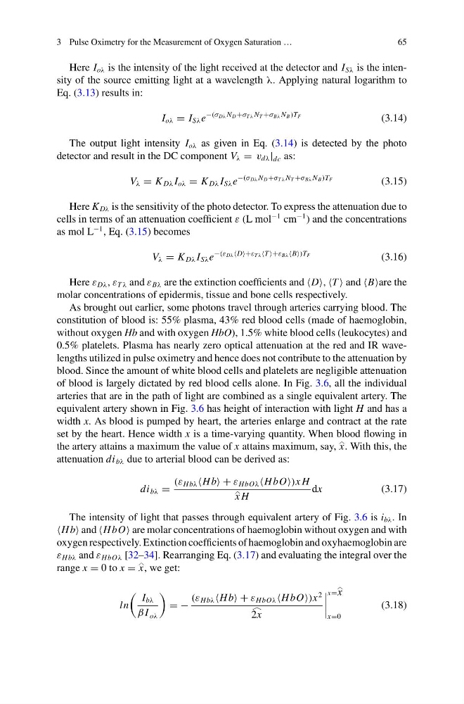
Here I oλ is the intensity of the light received at the detector and I Sλ is the intensity of the source emitting light at a wavelength λ. Applying natural logarithm to
Eq. (3.13) results in:
I oλ = I Sλ e
−(σ Dλ N D +σ T λ N T +σ Bλ N B )TF
(3.14)
The output light intensity I oλ as given in Eq. (3.14) is detected by the photo
detector and result in the DC component V λ = v dλ | dc as:
V λ = K Dλ I oλ = K Dλ I Sλ e
−(σ Dλ N D +σ T λ N T +σ Bλ N B )TF
(3.15)
Here K Dλ is the sensitivity of the photo detector. To express the attenuation due to
cells in terms of an attenuation coefficient ε (L mol
−1 cm
−1 ) and the concentrations
as mol L
−1 , Eq. (3.15) becomes
V λ = K Dλ I Sλ e
−(ε Dλ D+ε T λ T +ε Bλ B)T F
(3.16)
Here ε Dλ , ε T λ and ε Bλ are the extinction coefficients and D, T and Bare the
molar concentrations of epidermis, tissue and bone cells respectively.
As brought out earlier, some photons travel through arteries carrying blood. The
constitution of blood is: 55% plasma, 43% red blood cells (made of haemoglobin,
without oxygen Hb and with oxygen HbO), 1.5% white blood cells (leukocytes) and
0.5% platelets. Plasma has nearly zero optical attenuation at the red and IR wavelengths utilized in pulse oximetry and hence does not contribute to the attenuation by
blood. Since the amount of white blood cells and platelets are negligible attenuation
of blood is largely dictated by red blood cells alone. In Fig. 3.6, all the individual
arteries that are in the path of light are combined as a single equivalent artery. The
equivalent artery shown in Fig. 3.6 has height of interaction with light H and has a
width x. As blood is pumped by heart, the arteries enlarge and contract at the rate
set by the heart. Hence width x is a time-varying quantity. When blood flowing in
the artery attains a maximum the value of x attains maximum, say, x
. With this, the
attenuation di bλ due to arterial blood can be derived as:
di bλ =
(ε Hbλ Hb + ε HbOλ HbO)x H
x
H
dx
(3.17)
The intensity of light that passes through equivalent artery of Fig. 3.6 is i bλ . In
Hb and HbO are molar concentrations of haemoglobin without oxygen and with
oxygen respectively. Extinction coefficients of haemoglobin and oxyhaemoglobin are
ε Hbλ and ε HbOλ [32–34]. Rearranging Eq. (3.17) and evaluating the integral over the
range x = 0 to x = x
, we get:
ln
I bλ
β I oλ
= −
(ε Hbλ Hb + ε HbOλ HbO)x
2
2x
x=x
x=0
(3.18)
65
Here I oλ is the intensity of the light received at the detector and I Sλ is the intensity of the source emitting light at a wavelength λ. Applying natural logarithm to
Eq. (3.13) results in:
I oλ = I Sλ e
−(σ Dλ N D +σ T λ N T +σ Bλ N B )TF
(3.14)
The output light intensity I oλ as given in Eq. (3.14) is detected by the photo
detector and result in the DC component V λ = v dλ | dc as:
V λ = K Dλ I oλ = K Dλ I Sλ e
−(σ Dλ N D +σ T λ N T +σ Bλ N B )TF
(3.15)
Here K Dλ is the sensitivity of the photo detector. To express the attenuation due to
cells in terms of an attenuation coefficient ε (L mol
−1 cm
−1 ) and the concentrations
as mol L
−1 , Eq. (3.15) becomes
V λ = K Dλ I Sλ e
−(ε Dλ D+ε T λ T +ε Bλ B)T F
(3.16)
Here ε Dλ , ε T λ and ε Bλ are the extinction coefficients and D, T and Bare the
molar concentrations of epidermis, tissue and bone cells respectively.
As brought out earlier, some photons travel through arteries carrying blood. The
constitution of blood is: 55% plasma, 43% red blood cells (made of haemoglobin,
without oxygen Hb and with oxygen HbO), 1.5% white blood cells (leukocytes) and
0.5% platelets. Plasma has nearly zero optical attenuation at the red and IR wavelengths utilized in pulse oximetry and hence does not contribute to the attenuation by
blood. Since the amount of white blood cells and platelets are negligible attenuation
of blood is largely dictated by red blood cells alone. In Fig. 3.6, all the individual
arteries that are in the path of light are combined as a single equivalent artery. The
equivalent artery shown in Fig. 3.6 has height of interaction with light H and has a
width x. As blood is pumped by heart, the arteries enlarge and contract at the rate
set by the heart. Hence width x is a time-varying quantity. When blood flowing in
the artery attains a maximum the value of x attains maximum, say, x
. With this, the
attenuation di bλ due to arterial blood can be derived as:
di bλ =
(ε Hbλ Hb + ε HbOλ HbO)x H
x
H
dx
(3.17)
The intensity of light that passes through equivalent artery of Fig. 3.6 is i bλ . In
Hb and HbO are molar concentrations of haemoglobin without oxygen and with
oxygen respectively. Extinction coefficients of haemoglobin and oxyhaemoglobin are
ε Hbλ and ε HbOλ [32–34]. Rearranging Eq. (3.17) and evaluating the integral over the
range x = 0 to x = x
, we get:
ln
I bλ
β I oλ
= −
(ε Hbλ Hb + ε HbOλ HbO)x
2
2x
x=x
x=0
(3.18)