

44
M. Mukunda Rao
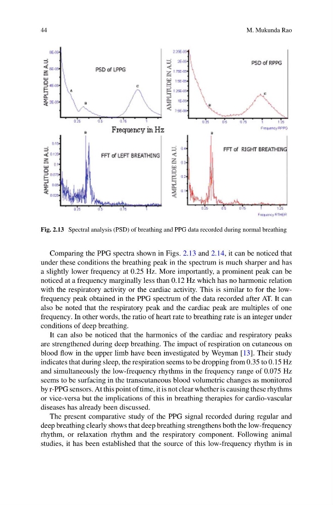
Fig. 2.13 Spectral analysis (PSD) of breathing and PPG data recorded during normal breathing
Comparing the PPG spectra shown in Figs. 2.13 and 2.14, it can be noticed that
under these conditions the breathing peak in the spectrum is much sharper and has
a slightly lower frequency at 0.25 Hz. More importantly, a prominent peak can be
noticed at a frequency marginally less than 0.12 Hz which has no harmonic relation
with the respiratory activity or the cardiac activity. This is similar to for the lowfrequency peak obtained in the PPG spectrum of the data recorded after AT. It can
also be noted that the respiratory peak and the cardiac peak are multiples of one
frequency. In other words, the ratio of heart rate to breathing rate is an integer under
conditions of deep breathing.
It can also be noticed that the harmonics of the cardiac and respiratory peaks
are strengthened during deep breathing. The impact of respiration on cutaneous on
blood flow in the upper limb have been investigated by Weyman [13]. Their study
indicates that during sleep, the respiration seems to be dropping from 0.35 to 0.15 Hz
and simultaneously the low-frequency rhythms in the frequency range of 0.075 Hz
seems to be surfacing in the transcutaneous blood volumetric changes as monitored
by r-PPG sensors. At this point of time, it is not clear whether is causing these rhythms
or vice-versa but the implications of this in breathing therapies for cardio-vascular
diseases has already been discussed.
The present comparative study of the PPG signal recorded during regular and
deep breathing clearly shows that deep breathing strengthens both the low-frequency
rhythm, or relaxation rhythm and the respiratory component. Following animal
studies, it has been established that the source of this low-frequency rhythm is in
M. Mukunda Rao
Fig. 2.13 Spectral analysis (PSD) of breathing and PPG data recorded during normal breathing
Comparing the PPG spectra shown in Figs. 2.13 and 2.14, it can be noticed that
under these conditions the breathing peak in the spectrum is much sharper and has
a slightly lower frequency at 0.25 Hz. More importantly, a prominent peak can be
noticed at a frequency marginally less than 0.12 Hz which has no harmonic relation
with the respiratory activity or the cardiac activity. This is similar to for the lowfrequency peak obtained in the PPG spectrum of the data recorded after AT. It can
also be noted that the respiratory peak and the cardiac peak are multiples of one
frequency. In other words, the ratio of heart rate to breathing rate is an integer under
conditions of deep breathing.
It can also be noticed that the harmonics of the cardiac and respiratory peaks
are strengthened during deep breathing. The impact of respiration on cutaneous on
blood flow in the upper limb have been investigated by Weyman [13]. Their study
indicates that during sleep, the respiration seems to be dropping from 0.35 to 0.15 Hz
and simultaneously the low-frequency rhythms in the frequency range of 0.075 Hz
seems to be surfacing in the transcutaneous blood volumetric changes as monitored
by r-PPG sensors. At this point of time, it is not clear whether is causing these rhythms
or vice-versa but the implications of this in breathing therapies for cardio-vascular
diseases has already been discussed.
The present comparative study of the PPG signal recorded during regular and
deep breathing clearly shows that deep breathing strengthens both the low-frequency
rhythm, or relaxation rhythm and the respiratory component. Following animal
studies, it has been established that the source of this low-frequency rhythm is in