

26
V. Blazek
others, the so called tissue perfusion index (TPI) and Oliva Roztocil index (ORI) as
extracted from the peripheral arterial pulse wave with suitable electronic filtering,
amplification and signal post-processing permits relevant insights into the peripheral
hemodynamical status of the monitored subject [61, 65, 66]. The slower (not heartsynchronous) rhythms in the dermal perfusion, as important as they are for the holistic
assessment of complex local perfusion dynamics, first remain unconsidered here.
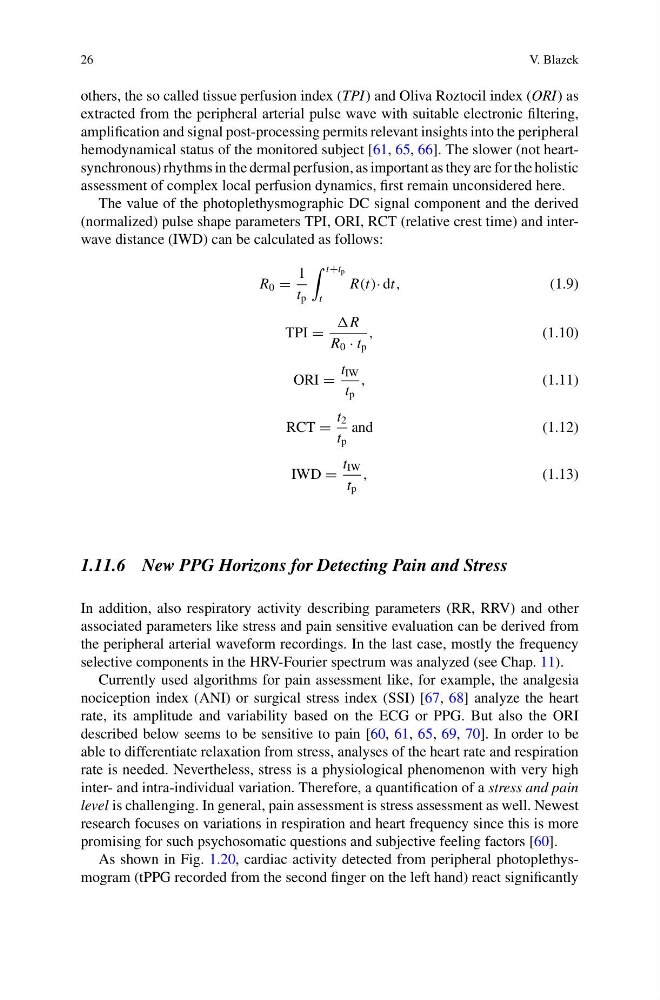
The value of the photoplethysmographic DC signal component and the derived
(normalized) pulse shape parameters TPI, ORI, RCT (relative crest time) and interwave distance (IWD) can be calculated as follows:
R 0 =
1
t p
t+t p
t
R(t)· dt,
(1.9)
TPI =
R
R 0 · t p
,
(1.10)
ORI =
t IW
t p
,
(1.11)
RCT =
t 2
t p
and
(1.12)
IWD =
t IW
t p
,
(1.13)
1.11.6 New PPG Horizons for Detecting Pain and Stress
In addition, also respiratory activity describing parameters (RR, RRV) and other
associated parameters like stress and pain sensitive evaluation can be derived from
the peripheral arterial waveform recordings. In the last case, mostly the frequency
selective components in the HRV-Fourier spectrum was analyzed (see Chap. 11).
Currently used algorithms for pain assessment like, for example, the analgesia
nociception index (ANI) or surgical stress index (SSI) [67, 68] analyze the heart
rate, its amplitude and variability based on the ECG or PPG. But also the ORI
described below seems to be sensitive to pain [60, 61, 65, 69, 70]. In order to be
able to differentiate relaxation from stress, analyses of the heart rate and respiration
rate is needed. Nevertheless, stress is a physiological phenomenon with very high
inter- and intra-individual variation. Therefore, a quantification of a stress and pain
level is challenging. In general, pain assessment is stress assessment as well. Newest
research focuses on variations in respiration and heart frequency since this is more
promising for such psychosomatic questions and subjective feeling factors [60].
As shown in Fig. 1.20, cardiac activity detected from peripheral photoplethysmogram (tPPG recorded from the second finger on the left hand) react significantly
V. Blazek
others, the so called tissue perfusion index (TPI) and Oliva Roztocil index (ORI) as
extracted from the peripheral arterial pulse wave with suitable electronic filtering,
amplification and signal post-processing permits relevant insights into the peripheral
hemodynamical status of the monitored subject [61, 65, 66]. The slower (not heartsynchronous) rhythms in the dermal perfusion, as important as they are for the holistic
assessment of complex local perfusion dynamics, first remain unconsidered here.
The value of the photoplethysmographic DC signal component and the derived
(normalized) pulse shape parameters TPI, ORI, RCT (relative crest time) and interwave distance (IWD) can be calculated as follows:
R 0 =
1
t p
t+t p
t
R(t)· dt,
(1.9)
TPI =
R
R 0 · t p
,
(1.10)
ORI =
t IW
t p
,
(1.11)
RCT =
t 2
t p
and
(1.12)
IWD =
t IW
t p
,
(1.13)
1.11.6 New PPG Horizons for Detecting Pain and Stress
In addition, also respiratory activity describing parameters (RR, RRV) and other
associated parameters like stress and pain sensitive evaluation can be derived from
the peripheral arterial waveform recordings. In the last case, mostly the frequency
selective components in the HRV-Fourier spectrum was analyzed (see Chap. 11).
Currently used algorithms for pain assessment like, for example, the analgesia
nociception index (ANI) or surgical stress index (SSI) [67, 68] analyze the heart
rate, its amplitude and variability based on the ECG or PPG. But also the ORI
described below seems to be sensitive to pain [60, 61, 65, 69, 70]. In order to be
able to differentiate relaxation from stress, analyses of the heart rate and respiration
rate is needed. Nevertheless, stress is a physiological phenomenon with very high
inter- and intra-individual variation. Therefore, a quantification of a stress and pain
level is challenging. In general, pain assessment is stress assessment as well. Newest
research focuses on variations in respiration and heart frequency since this is more
promising for such psychosomatic questions and subjective feeling factors [60].
As shown in Fig. 1.20, cardiac activity detected from peripheral photoplethysmogram (tPPG recorded from the second finger on the left hand) react significantly