

1 Skin Perfusion Studies: Historical Notes and Modern Measuring …
27
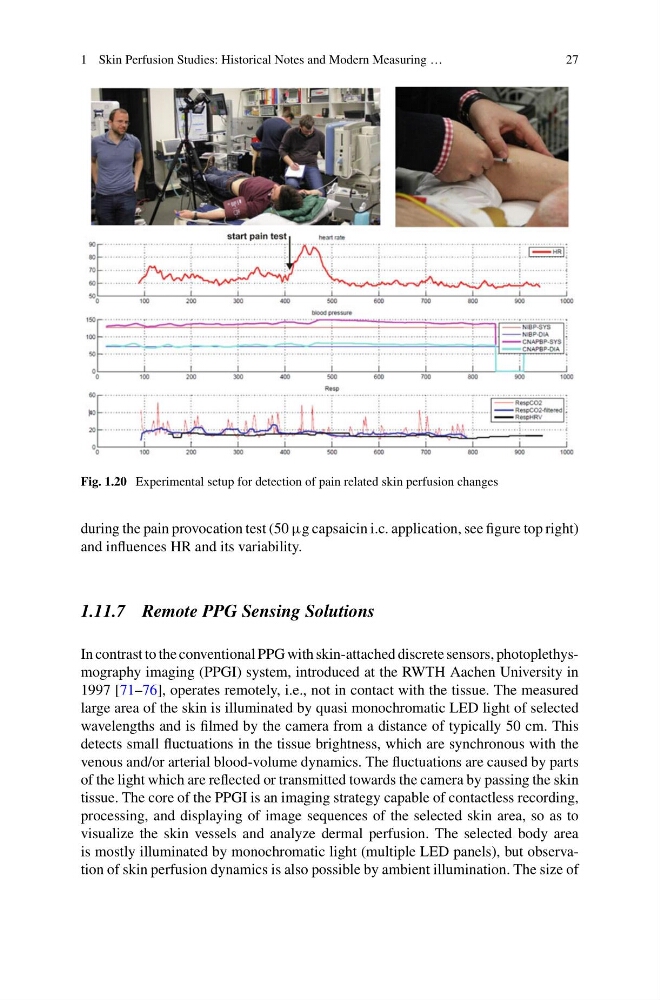
Fig. 1.20 Experimental setup for detection of pain related skin perfusion changes
during the pain provocation test (50 µg capsaicin i.c. application, see figure top right)
and influences HR and its variability.
1.11.7 Remote PPG Sensing Solutions
In contrast to the conventional PPG with skin-attached discrete sensors, photoplethysmography imaging (PPGI) system, introduced at the RWTH Aachen University in
1997 [71–76], operates remotely, i.e., not in contact with the tissue. The measured
large area of the skin is illuminated by quasi monochromatic LED light of selected
wavelengths and is filmed by the camera from a distance of typically 50 cm. This
detects small fluctuations in the tissue brightness, which are synchronous with the
venous and/or arterial blood-volume dynamics. The fluctuations are caused by parts
of the light which are reflected or transmitted towards the camera by passing the skin
tissue. The core of the PPGI is an imaging strategy capable of contactless recording,
processing, and displaying of image sequences of the selected skin area, so as to
visualize the skin vessels and analyze dermal perfusion. The selected body area
is mostly illuminated by monochromatic light (multiple LED panels), but observation of skin perfusion dynamics is also possible by ambient illumination. The size of
27
Fig. 1.20 Experimental setup for detection of pain related skin perfusion changes
during the pain provocation test (50 µg capsaicin i.c. application, see figure top right)
and influences HR and its variability.
1.11.7 Remote PPG Sensing Solutions
In contrast to the conventional PPG with skin-attached discrete sensors, photoplethysmography imaging (PPGI) system, introduced at the RWTH Aachen University in
1997 [71–76], operates remotely, i.e., not in contact with the tissue. The measured
large area of the skin is illuminated by quasi monochromatic LED light of selected
wavelengths and is filmed by the camera from a distance of typically 50 cm. This
detects small fluctuations in the tissue brightness, which are synchronous with the
venous and/or arterial blood-volume dynamics. The fluctuations are caused by parts
of the light which are reflected or transmitted towards the camera by passing the skin
tissue. The core of the PPGI is an imaging strategy capable of contactless recording,
processing, and displaying of image sequences of the selected skin area, so as to
visualize the skin vessels and analyze dermal perfusion. The selected body area
is mostly illuminated by monochromatic light (multiple LED panels), but observation of skin perfusion dynamics is also possible by ambient illumination. The size of