

1 Skin Perfusion Studies: Historical Notes and Modern Measuring …
25
investigated volume of blood. Pulse oximetry devices that usually detect the heart
synchronous arterial blood volume pulse on fingers and toes (more recently, in the
ear canal) is nowadays present in any ambulances and on any intensive care unit;
however, they are used with equal success for long-term monitoring of serious injuries
and patients with hemodynamic instability, increased cardiovascular risk, respiratory
failure and other risk conditions.
The venous oxygen saturation might recently be calculated from peripheral venous
blood volume fluctuations that occur in a specific sequence leg exercise (producing
so called venous pulse). Since the arterial oxygen saturation is calculated too, the
local oxygen consumption can be determined. Normally, the arterial blood saturation
value SpO 2 should be around 98% and the peripheral venous saturation (SvO 2 ) value
at about 78%, so that the peripheral local saturation consumption is 20% in this case
(see Chap. 5).
1.11.5 Analyzing Pulsatile Component of the PPG Waveform
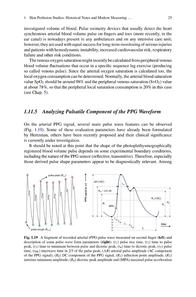
On the arterial PPG signal, several main pulse wave features can be observed
(Fig. 1.19). Some of these evaluation parameters have already been formulated
by Hertzman, others have been recently proposed and their clinical significance
is currently under investigation.
It should be noted at this point that the shape of the photoplethysmographically
registered blood volume pulse depends on some experimental boundary conditions,
including the nature of the PPG sensor (reflective, transmitive). Therefore, especially
those derived pulse shape parameters appear to be diagnostically relevant. Among
Fig. 1.19 A fragment of recorded arterial rPPG pulse wave measured on second finger (left) and
description of some pulse wave form parameters (right): (t 1 ) pulse rise time, (t 2 ) time to pulse
peak, (t 3 ) time to minimum between pulse and dicrotic peak, (t 4 ) time to dicrotic peak, (t 5 ) pulse
time, (t IW ) interwave time in 2/3 of the pulse peak, (R) arterial pulse amplitude (AC component
of the PPG signal), (R 0 ) DC component of the PPG signal, (R 1 ) inflection point amplitude, (R 3 )
interims minimum amplitude, (R 4 ) dicrotic peak amplitude and (MPA) maximal pulse acceleration
25
investigated volume of blood. Pulse oximetry devices that usually detect the heart
synchronous arterial blood volume pulse on fingers and toes (more recently, in the
ear canal) is nowadays present in any ambulances and on any intensive care unit;
however, they are used with equal success for long-term monitoring of serious injuries
and patients with hemodynamic instability, increased cardiovascular risk, respiratory
failure and other risk conditions.
The venous oxygen saturation might recently be calculated from peripheral venous
blood volume fluctuations that occur in a specific sequence leg exercise (producing
so called venous pulse). Since the arterial oxygen saturation is calculated too, the
local oxygen consumption can be determined. Normally, the arterial blood saturation
value SpO 2 should be around 98% and the peripheral venous saturation (SvO 2 ) value
at about 78%, so that the peripheral local saturation consumption is 20% in this case
(see Chap. 5).
1.11.5 Analyzing Pulsatile Component of the PPG Waveform
On the arterial PPG signal, several main pulse wave features can be observed
(Fig. 1.19). Some of these evaluation parameters have already been formulated
by Hertzman, others have been recently proposed and their clinical significance
is currently under investigation.
It should be noted at this point that the shape of the photoplethysmographically
registered blood volume pulse depends on some experimental boundary conditions,
including the nature of the PPG sensor (reflective, transmitive). Therefore, especially
those derived pulse shape parameters appear to be diagnostically relevant. Among
Fig. 1.19 A fragment of recorded arterial rPPG pulse wave measured on second finger (left) and
description of some pulse wave form parameters (right): (t 1 ) pulse rise time, (t 2 ) time to pulse
peak, (t 3 ) time to minimum between pulse and dicrotic peak, (t 4 ) time to dicrotic peak, (t 5 ) pulse
time, (t IW ) interwave time in 2/3 of the pulse peak, (R) arterial pulse amplitude (AC component
of the PPG signal), (R 0 ) DC component of the PPG signal, (R 1 ) inflection point amplitude, (R 3 )
interims minimum amplitude, (R 4 ) dicrotic peak amplitude and (MPA) maximal pulse acceleration