

16
V. Blazek
to a computer after A/D conversion to visualize and store these data. With our
measurement setup all functional settings and sensor controls can be managed via
PC.
1.9 Monitoring of Skin Perfusion Dynamics Under
Controlled Conditions in Time and Frequency Domain
This kind of post processing of PPG signals was first implemented and published in
1985 with the aim to analyze pulsatile perfusion signals in both time and frequency
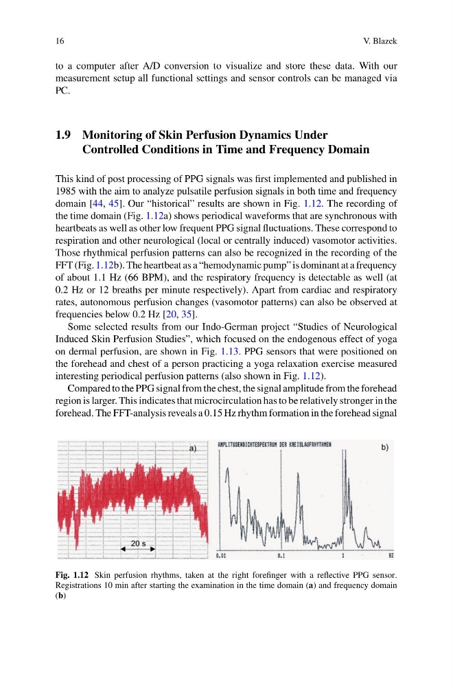
domain [44, 45]. Our “historical” results are shown in Fig. 1.12. The recording of
the time domain (Fig. 1.12a) shows periodical waveforms that are synchronous with
heartbeats as well as other low frequent PPG signal fluctuations. These correspond to
respiration and other neurological (local or centrally induced) vasomotor activities.
Those rhythmical perfusion patterns can also be recognized in the recording of the
FFT (Fig. 1.12b). The heartbeat as a “hemodynamic pump” is dominant at a frequency
of about 1.1 Hz (66 BPM), and the respiratory frequency is detectable as well (at
0.2 Hz or 12 breaths per minute respectively). Apart from cardiac and respiratory
rates, autonomous perfusion changes (vasomotor patterns) can also be observed at
frequencies below 0.2 Hz [20, 35].
Some selected results from our Indo-German project “Studies of Neurological
Induced Skin Perfusion Studies”, which focused on the endogenous effect of yoga
on dermal perfusion, are shown in Fig. 1.13. PPG sensors that were positioned on
the forehead and chest of a person practicing a yoga relaxation exercise measured
interesting periodical perfusion patterns (also shown in Fig. 1.12).
Compared to the PPG signal from the chest, the signal amplitude from the forehead
region is larger. This indicates that microcirculation has to be relatively stronger in the
forehead. The FFT-analysis reveals a 0.15 Hz rhythm formation in the forehead signal
Fig. 1.12 Skin perfusion rhythms, taken at the right forefinger with a reflective PPG sensor.
Registrations 10 min after starting the examination in the time domain (a) and frequency domain
(b)
V. Blazek
to a computer after A/D conversion to visualize and store these data. With our
measurement setup all functional settings and sensor controls can be managed via
PC.
1.9 Monitoring of Skin Perfusion Dynamics Under
Controlled Conditions in Time and Frequency Domain
This kind of post processing of PPG signals was first implemented and published in
1985 with the aim to analyze pulsatile perfusion signals in both time and frequency
domain [44, 45]. Our “historical” results are shown in Fig. 1.12. The recording of
the time domain (Fig. 1.12a) shows periodical waveforms that are synchronous with
heartbeats as well as other low frequent PPG signal fluctuations. These correspond to
respiration and other neurological (local or centrally induced) vasomotor activities.
Those rhythmical perfusion patterns can also be recognized in the recording of the
FFT (Fig. 1.12b). The heartbeat as a “hemodynamic pump” is dominant at a frequency
of about 1.1 Hz (66 BPM), and the respiratory frequency is detectable as well (at
0.2 Hz or 12 breaths per minute respectively). Apart from cardiac and respiratory
rates, autonomous perfusion changes (vasomotor patterns) can also be observed at
frequencies below 0.2 Hz [20, 35].
Some selected results from our Indo-German project “Studies of Neurological
Induced Skin Perfusion Studies”, which focused on the endogenous effect of yoga
on dermal perfusion, are shown in Fig. 1.13. PPG sensors that were positioned on
the forehead and chest of a person practicing a yoga relaxation exercise measured
interesting periodical perfusion patterns (also shown in Fig. 1.12).
Compared to the PPG signal from the chest, the signal amplitude from the forehead
region is larger. This indicates that microcirculation has to be relatively stronger in the
forehead. The FFT-analysis reveals a 0.15 Hz rhythm formation in the forehead signal
Fig. 1.12 Skin perfusion rhythms, taken at the right forefinger with a reflective PPG sensor.
Registrations 10 min after starting the examination in the time domain (a) and frequency domain
(b)