

10
V. Blazek
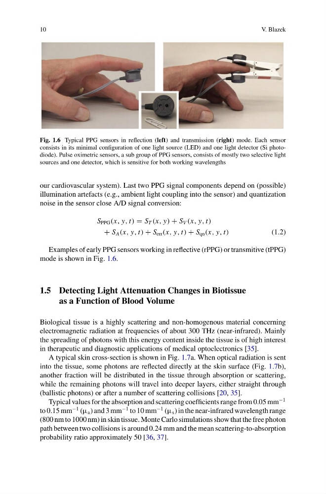
Fig. 1.6 Typical PPG sensors in reflection (left) and transmission (right) mode. Each sensor
consists in its minimal configuration of one light source (LED) and one light detector (Si photodiode). Pulse oximetric sensors, a sub group of PPG sensors, consists of mostly two selective light
sources and one detector, which is sensitive for both working wavelengths
our cardiovascular system). Last two PPG signal components depend on (possible)
illumination artefacts (e.g., ambient light coupling into the sensor) and quantization
noise in the sensor close A/D signal conversion:
S PPG (x, y, t) = S T (x, y) + S V (x, y, t)
+ S A (x, y, t) + S err (x, y, t) + S qn (x, y, t)
(1.2)
Examples of early PPG sensors working in reflective (rPPG) or transmitive (tPPG)
mode is shown in Fig. 1.6.
1.5 Detecting Light Attenuation Changes in Biotissue
as a Function of Blood Volume
Biological tissue is a highly scattering and non-homogenous material concerning
electromagnetic radiation at frequencies of about 300 THz (near-infrared). Mainly
the spreading of photons with this energy content inside the tissue is of high interest
in therapeutic and diagnostic applications of medical optoelectronics [35].
A typical skin cross-section is shown in Fig. 1.7a. When optical radiation is sent
into the tissue, some photons are reflected directly at the skin surface (Fig. 1.7b),
another fraction will be distributed in the tissue through absorption or scattering,
while the remaining photons will travel into deeper layers, either straight through
(ballistic photons) or after a number of scattering collisions [20, 35].
Typical values for the absorption and scattering coefficients range from 0.05 mm
−1
to 0.15 mm
−1 (µ a ) and 3 mm
−1 to 10 mm
−1 (µ s ) in the near-infrared wavelength range
(800 nm to 1000 nm) in skin tissue. Monte Carlo simulations show that the free photon
path between two collisions is around 0.24 mm and the mean scattering-to-absorption
probability ratio approximately 50 [36, 37].
V. Blazek
Fig. 1.6 Typical PPG sensors in reflection (left) and transmission (right) mode. Each sensor
consists in its minimal configuration of one light source (LED) and one light detector (Si photodiode). Pulse oximetric sensors, a sub group of PPG sensors, consists of mostly two selective light
sources and one detector, which is sensitive for both working wavelengths
our cardiovascular system). Last two PPG signal components depend on (possible)
illumination artefacts (e.g., ambient light coupling into the sensor) and quantization
noise in the sensor close A/D signal conversion:
S PPG (x, y, t) = S T (x, y) + S V (x, y, t)
+ S A (x, y, t) + S err (x, y, t) + S qn (x, y, t)
(1.2)
Examples of early PPG sensors working in reflective (rPPG) or transmitive (tPPG)
mode is shown in Fig. 1.6.
1.5 Detecting Light Attenuation Changes in Biotissue
as a Function of Blood Volume
Biological tissue is a highly scattering and non-homogenous material concerning
electromagnetic radiation at frequencies of about 300 THz (near-infrared). Mainly
the spreading of photons with this energy content inside the tissue is of high interest
in therapeutic and diagnostic applications of medical optoelectronics [35].
A typical skin cross-section is shown in Fig. 1.7a. When optical radiation is sent
into the tissue, some photons are reflected directly at the skin surface (Fig. 1.7b),
another fraction will be distributed in the tissue through absorption or scattering,
while the remaining photons will travel into deeper layers, either straight through
(ballistic photons) or after a number of scattering collisions [20, 35].
Typical values for the absorption and scattering coefficients range from 0.05 mm
−1
to 0.15 mm
−1 (µ a ) and 3 mm
−1 to 10 mm
−1 (µ s ) in the near-infrared wavelength range
(800 nm to 1000 nm) in skin tissue. Monte Carlo simulations show that the free photon
path between two collisions is around 0.24 mm and the mean scattering-to-absorption
probability ratio approximately 50 [36, 37].