

1 Skin Perfusion Studies: Historical Notes and Modern Measuring …
9
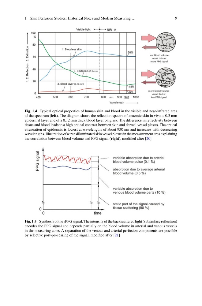
Fig. 1.4 Typical optical properties of human skin and blood in the visible and near-infrared area
of the spectrum (left). The diagram shows the reflection spectra of anaemic skin in vivo, a 0.3 mm
epidermal layer and of a 0.12 mm thick blood layer on glass. The difference in reflectivity between
tissue and blood leads to a high optical contrast between skin and dermal vessel plexus. The optical
attenuation of epidermis is lowest at wavelengths of about 930 nm and increases with decreasing
wavelengths. Illustration of a transilluminated skin vessel plexus in the measurement area explaining
the correlation between blood volume and PPG signal (right), modified after [20]
Fig. 1.5 Synthesis of the rPPG signal. The intensity of the backscattered light (subsurface reflection)
encodes the PPG signal and depends partially on the blood volume in arterial and venous vessels
in the measuring zone. A separation of the venous and arterial perfusion components are possible
by selective post-processing of the signal, modified after [21]
9
Fig. 1.4 Typical optical properties of human skin and blood in the visible and near-infrared area
of the spectrum (left). The diagram shows the reflection spectra of anaemic skin in vivo, a 0.3 mm
epidermal layer and of a 0.12 mm thick blood layer on glass. The difference in reflectivity between
tissue and blood leads to a high optical contrast between skin and dermal vessel plexus. The optical
attenuation of epidermis is lowest at wavelengths of about 930 nm and increases with decreasing
wavelengths. Illustration of a transilluminated skin vessel plexus in the measurement area explaining
the correlation between blood volume and PPG signal (right), modified after [20]
Fig. 1.5 Synthesis of the rPPG signal. The intensity of the backscattered light (subsurface reflection)
encodes the PPG signal and depends partially on the blood volume in arterial and venous vessels
in the measuring zone. A separation of the venous and arterial perfusion components are possible
by selective post-processing of the signal, modified after [21]