

8
V. Blazek
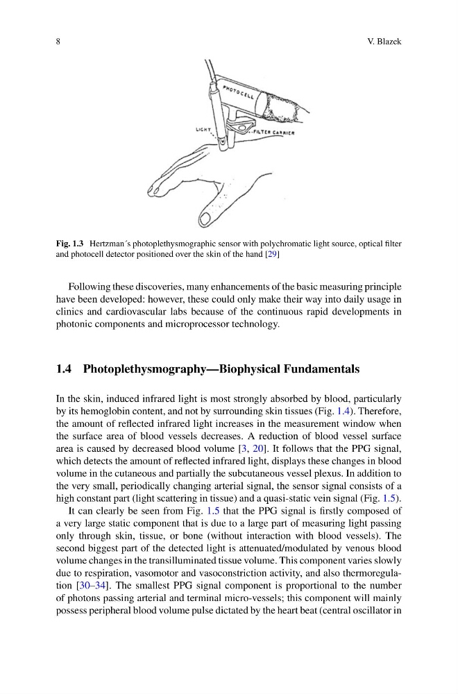
Fig. 1.3 Hertzman´s photoplethysmographic sensor with polychromatic light source, optical filter
and photocell detector positioned over the skin of the hand [29]
Following these discoveries, many enhancements of the basic measuring principle
have been developed: however, these could only make their way into daily usage in
clinics and cardiovascular labs because of the continuous rapid developments in
photonic components and microprocessor technology.
1.4 Photoplethysmography—Biophysical Fundamentals
In the skin, induced infrared light is most strongly absorbed by blood, particularly
by its hemoglobin content, and not by surrounding skin tissues (Fig. 1.4). Therefore,
the amount of reflected infrared light increases in the measurement window when
the surface area of blood vessels decreases. A reduction of blood vessel surface
area is caused by decreased blood volume [3, 20]. It follows that the PPG signal,
which detects the amount of reflected infrared light, displays these changes in blood
volume in the cutaneous and partially the subcutaneous vessel plexus. In addition to
the very small, periodically changing arterial signal, the sensor signal consists of a
high constant part (light scattering in tissue) and a quasi-static vein signal (Fig. 1.5).
It can clearly be seen from Fig. 1.5 that the PPG signal is firstly composed of
a very large static component that is due to a large part of measuring light passing
only through skin, tissue, or bone (without interaction with blood vessels). The
second biggest part of the detected light is attenuated/modulated by venous blood
volume changes in the transilluminated tissue volume. This component varies slowly
due to respiration, vasomotor and vasoconstriction activity, and also thermoregulation [30–34]. The smallest PPG signal component is proportional to the number
of photons passing arterial and terminal micro-vessels; this component will mainly
possess peripheral blood volume pulse dictated by the heart beat (central oscillator in
V. Blazek
Fig. 1.3 Hertzman´s photoplethysmographic sensor with polychromatic light source, optical filter
and photocell detector positioned over the skin of the hand [29]
Following these discoveries, many enhancements of the basic measuring principle
have been developed: however, these could only make their way into daily usage in
clinics and cardiovascular labs because of the continuous rapid developments in
photonic components and microprocessor technology.
1.4 Photoplethysmography—Biophysical Fundamentals
In the skin, induced infrared light is most strongly absorbed by blood, particularly
by its hemoglobin content, and not by surrounding skin tissues (Fig. 1.4). Therefore,
the amount of reflected infrared light increases in the measurement window when
the surface area of blood vessels decreases. A reduction of blood vessel surface
area is caused by decreased blood volume [3, 20]. It follows that the PPG signal,
which detects the amount of reflected infrared light, displays these changes in blood
volume in the cutaneous and partially the subcutaneous vessel plexus. In addition to
the very small, periodically changing arterial signal, the sensor signal consists of a
high constant part (light scattering in tissue) and a quasi-static vein signal (Fig. 1.5).
It can clearly be seen from Fig. 1.5 that the PPG signal is firstly composed of
a very large static component that is due to a large part of measuring light passing
only through skin, tissue, or bone (without interaction with blood vessels). The
second biggest part of the detected light is attenuated/modulated by venous blood
volume changes in the transilluminated tissue volume. This component varies slowly
due to respiration, vasomotor and vasoconstriction activity, and also thermoregulation [30–34]. The smallest PPG signal component is proportional to the number
of photons passing arterial and terminal micro-vessels; this component will mainly
possess peripheral blood volume pulse dictated by the heart beat (central oscillator in