

1 Skin Perfusion Studies: Historical Notes and Modern Measuring …
11
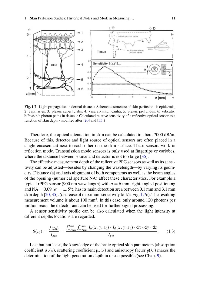
Fig. 1.7 Light propagation in dermal tissue. a Schematic structure of skin perfusion. 1: epidermis,
2: capillaries, 3: plexus superficialis, 4: vasa communicantia, 5: plexus profundus, 6: subcutis.
b Possible photon paths in tissue. c Calculated relative sensitivity of a reflective optical sensor as a
function of skin depth (modified after [20] and [35])
Therefore, the optical attenuation in skin can be calculated to about 7000 dB/m.
Because of this, detector and light source of optical sensors are often placed in a
single encasement next to each other on the skin surface. These sensors work in
reflection mode. Transmission mode sensors is only used at fingertips or earlobes,
where the distance between source and detector is not too large [35].
The effective measurement depth of the reflective PPG sensors as well as its sensitivity can be adjusted—besides by changing the wavelength—by varying its geometry. Distance (a) and axis alignment of both components as well as the beam angles
of the opening (numerical aperture NA) affect these characteristics. For example a
typical rPPG sensor (900 nm wavelength) with a = 6 mm, right-angled positioning
and NA = 0.09 (α = ± 5°), has its main detection area between 0.1 mm and 3.1 mm
skin depth [20, 35]. (decrease of maximum sensitivity to 1/e, Fig. 1.7c). The resulting
measurement volume is about 100 mm
3 . In this case, only around 120 photons per
million reach the detector and can be used for further signal processing.
A sensor sensitivity profile can be also calculated when the light intensity at
different depths locations are regarded.
S(z 0 ) =
I (z 0 )
I ges
=
y max
−y max
x max
−x max
I q (x, y, z 0 ) · I d (x, y, z 0 ) · dx · dy · dz
I ges
.
(1.3)
Last but not least, the knowledge of the basic optical skin parameters (absorption
coefficient µ a (λ), scattering coefficient µ s (λ) and anisotropy factor g(λ)) makes the
determination of the light penetration depth in tissue possible (see Chap. 9).
11
Fig. 1.7 Light propagation in dermal tissue. a Schematic structure of skin perfusion. 1: epidermis,
2: capillaries, 3: plexus superficialis, 4: vasa communicantia, 5: plexus profundus, 6: subcutis.
b Possible photon paths in tissue. c Calculated relative sensitivity of a reflective optical sensor as a
function of skin depth (modified after [20] and [35])
Therefore, the optical attenuation in skin can be calculated to about 7000 dB/m.
Because of this, detector and light source of optical sensors are often placed in a
single encasement next to each other on the skin surface. These sensors work in
reflection mode. Transmission mode sensors is only used at fingertips or earlobes,
where the distance between source and detector is not too large [35].
The effective measurement depth of the reflective PPG sensors as well as its sensitivity can be adjusted—besides by changing the wavelength—by varying its geometry. Distance (a) and axis alignment of both components as well as the beam angles
of the opening (numerical aperture NA) affect these characteristics. For example a
typical rPPG sensor (900 nm wavelength) with a = 6 mm, right-angled positioning
and NA = 0.09 (α = ± 5°), has its main detection area between 0.1 mm and 3.1 mm
skin depth [20, 35]. (decrease of maximum sensitivity to 1/e, Fig. 1.7c). The resulting
measurement volume is about 100 mm
3 . In this case, only around 120 photons per
million reach the detector and can be used for further signal processing.
A sensor sensitivity profile can be also calculated when the light intensity at
different depths locations are regarded.
S(z 0 ) =
I (z 0 )
I ges
=
y max
−y max
x max
−x max
I q (x, y, z 0 ) · I d (x, y, z 0 ) · dx · dy · dz
I ges
.
(1.3)
Last but not least, the knowledge of the basic optical skin parameters (absorption
coefficient µ a (λ), scattering coefficient µ s (λ) and anisotropy factor g(λ)) makes the
determination of the light penetration depth in tissue possible (see Chap. 9).