

13 Concluding Remarks and New Horizons in Skin Perfusion Studies
225
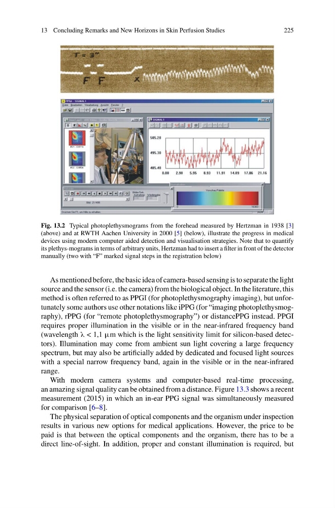
Fig. 13.2 Typical photoplethysmograms from the forehead measured by Hertzman in 1938 [3]
(above) and at RWTH Aachen University in 2000 [5] (below), illustrate the progress in medical
devices using modern computer aided detection and visualisation strategies. Note that to quantify
its plethys-mograms in terms of arbitrary units, Hertzman had to insert a filter in front of the detector
manually (two with “F” marked signal steps in the registration below)
As mentioned before, the basic idea of camera-based sensing is to separate the light
source and the sensor (i.e. the camera) from the biological object. In the literature, this
method is often referred to as PPGI (for photoplethysmography imaging), but unfortunately some authors use other notations like iPPG (for “imaging photoplethysmography), rPPG (for “remote photoplethysmography”) or distancePPG instead. PPGI
requires proper illumination in the visible or in the near-infrared frequency band
(wavelength λ < 1,1 μm which is the light sensitivity limit for silicon-based detectors). Illumination may come from ambient sun light covering a large frequency
spectrum, but may also be artificially added by dedicated and focused light sources
with a special narrow frequency band, again in the visible or in the near-infrared
range.
With modern camera systems and computer-based real-time processing,
an amazing signal quality can be obtained from a distance. Figure 13.3 shows a recent
measurement (2015) in which an in-ear PPG signal was simultaneously measured
for comparison [6–8].
The physical separation of optical components and the organism under inspection
results in various new options for medical applications. However, the price to be
paid is that between the optical components and the organism, there has to be a
direct line-of-sight. In addition, proper and constant illumination is required, but
225
Fig. 13.2 Typical photoplethysmograms from the forehead measured by Hertzman in 1938 [3]
(above) and at RWTH Aachen University in 2000 [5] (below), illustrate the progress in medical
devices using modern computer aided detection and visualisation strategies. Note that to quantify
its plethys-mograms in terms of arbitrary units, Hertzman had to insert a filter in front of the detector
manually (two with “F” marked signal steps in the registration below)
As mentioned before, the basic idea of camera-based sensing is to separate the light
source and the sensor (i.e. the camera) from the biological object. In the literature, this
method is often referred to as PPGI (for photoplethysmography imaging), but unfortunately some authors use other notations like iPPG (for “imaging photoplethysmography), rPPG (for “remote photoplethysmography”) or distancePPG instead. PPGI
requires proper illumination in the visible or in the near-infrared frequency band
(wavelength λ < 1,1 μm which is the light sensitivity limit for silicon-based detectors). Illumination may come from ambient sun light covering a large frequency
spectrum, but may also be artificially added by dedicated and focused light sources
with a special narrow frequency band, again in the visible or in the near-infrared
range.
With modern camera systems and computer-based real-time processing,
an amazing signal quality can be obtained from a distance. Figure 13.3 shows a recent
measurement (2015) in which an in-ear PPG signal was simultaneously measured
for comparison [6–8].
The physical separation of optical components and the organism under inspection
results in various new options for medical applications. However, the price to be
paid is that between the optical components and the organism, there has to be a
direct line-of-sight. In addition, proper and constant illumination is required, but