

224
S. Leonhardt
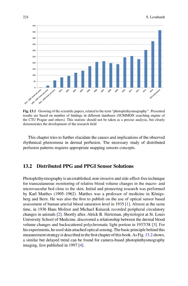
Fig. 13.1 Growing of the scientific papers, related to the term “photoplethysmography” . Presented
results are based on number of findings in different databases (SUMMON searching engine of
the CTU Prague and others). This statistic should not be taken as a precise analysis, but clearly
demonstrates the development of the research field
This chapter tries to further elucidate the causes and implications of the observed
rhythmical phenomena in dermal perfusion. The necessary study of distributed
perfusion patterns requires appropriate mapping sensors concepts.
13.2 Distributed PPG and PPGI Sensor Solutions
Photoplethysmography is an established, non-invasive and side-effect-free technique
for transcutaneous monitoring of relative blood volume changes in the macro- and
microvascular bed close to the skin. Initial and pioneering research was performed
by Karl Matthes (1905–1962). Matthes was a professor of medicine in Königsberg and Bern. He was also the first to publish on the use of optical sensor based
assessment of human arterial blood saturation level in 1935 [1]. Almost at the same
time, in 1936 Hans Molitor and Michael Kniazuk recorded peripheral circulatory
changes in animals [2]. Shortly after, Alrick B. Hertzman, physiologist at St. Louis
University School of Medicine, discovered a relationship between the dermal blood
volume changes and backscattered polychromatic light portion in 1937/38 [3]. For
his experiments, he used skin attached optical sensing. The basic principle behind this
measurement strategy is described in the first chapter of this book. As Fig. 13.2 shows,
a similar but delayed trend can be found for camera-based photoplethysmography
imaging, first published in 1997 [4].
S. Leonhardt
Fig. 13.1 Growing of the scientific papers, related to the term “photoplethysmography” . Presented
results are based on number of findings in different databases (SUMMON searching engine of
the CTU Prague and others). This statistic should not be taken as a precise analysis, but clearly
demonstrates the development of the research field
This chapter tries to further elucidate the causes and implications of the observed
rhythmical phenomena in dermal perfusion. The necessary study of distributed
perfusion patterns requires appropriate mapping sensors concepts.
13.2 Distributed PPG and PPGI Sensor Solutions
Photoplethysmography is an established, non-invasive and side-effect-free technique
for transcutaneous monitoring of relative blood volume changes in the macro- and
microvascular bed close to the skin. Initial and pioneering research was performed
by Karl Matthes (1905–1962). Matthes was a professor of medicine in Königsberg and Bern. He was also the first to publish on the use of optical sensor based
assessment of human arterial blood saturation level in 1935 [1]. Almost at the same
time, in 1936 Hans Molitor and Michael Kniazuk recorded peripheral circulatory
changes in animals [2]. Shortly after, Alrick B. Hertzman, physiologist at St. Louis
University School of Medicine, discovered a relationship between the dermal blood
volume changes and backscattered polychromatic light portion in 1937/38 [3]. For
his experiments, he used skin attached optical sensing. The basic principle behind this
measurement strategy is described in the first chapter of this book. As Fig. 13.2 shows,
a similar but delayed trend can be found for camera-based photoplethysmography
imaging, first published in 1997 [4].