

226
S. Leonhardt
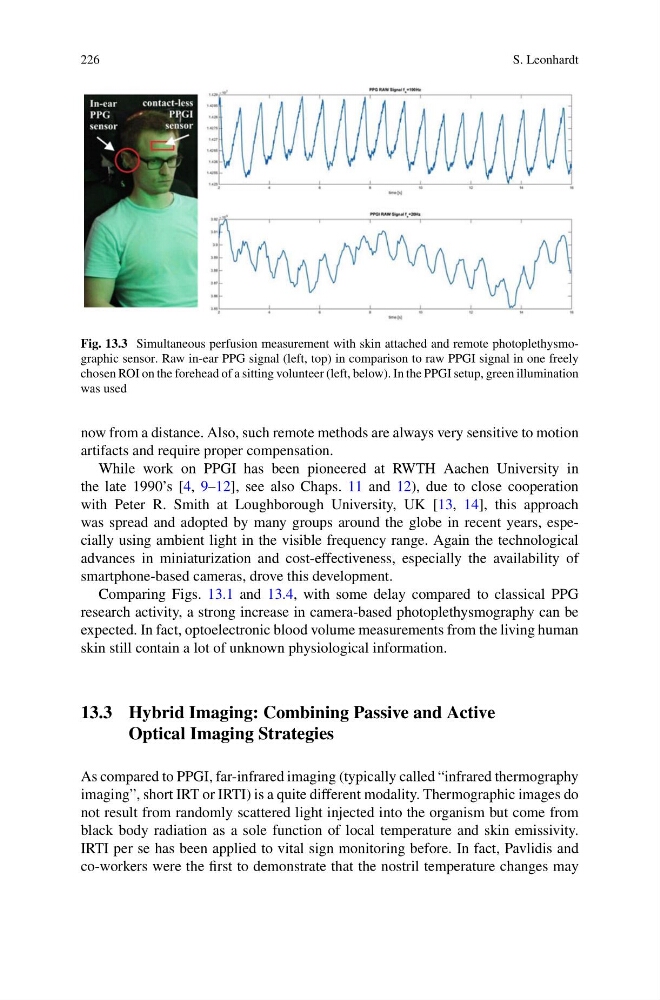
Fig. 13.3 Simultaneous perfusion measurement with skin attached and remote photoplethysmographic sensor. Raw in-ear PPG signal (left, top) in comparison to raw PPGI signal in one freely
chosen ROI on the forehead of a sitting volunteer (left, below). In the PPGI setup, green illumination
was used
now from a distance. Also, such remote methods are always very sensitive to motion
artifacts and require proper compensation.
While work on PPGI has been pioneered at RWTH Aachen University in
the late 1990’s [4, 9–12], see also Chaps. 11 and 12), due to close cooperation
with Peter R. Smith at Loughborough University, UK [13, 14], this approach
was spread and adopted by many groups around the globe in recent years, especially using ambient light in the visible frequency range. Again the technological
advances in miniaturization and cost-effectiveness, especially the availability of
smartphone-based cameras, drove this development.
Comparing Figs. 13.1 and 13.4, with some delay compared to classical PPG
research activity, a strong increase in camera-based photoplethysmography can be
expected. In fact, optoelectronic blood volume measurements from the living human
skin still contain a lot of unknown physiological information.
13.3 Hybrid Imaging: Combining Passive and Active
Optical Imaging Strategies
As compared to PPGI, far-infrared imaging (typically called “infrared thermography
imaging”, short IRT or IRTI) is a quite different modality. Thermographic images do
not result from randomly scattered light injected into the organism but come from
black body radiation as a sole function of local temperature and skin emissivity.
IRTI per se has been applied to vital sign monitoring before. In fact, Pavlidis and
co-workers were the first to demonstrate that the nostril temperature changes may
S. Leonhardt
Fig. 13.3 Simultaneous perfusion measurement with skin attached and remote photoplethysmographic sensor. Raw in-ear PPG signal (left, top) in comparison to raw PPGI signal in one freely
chosen ROI on the forehead of a sitting volunteer (left, below). In the PPGI setup, green illumination
was used
now from a distance. Also, such remote methods are always very sensitive to motion
artifacts and require proper compensation.
While work on PPGI has been pioneered at RWTH Aachen University in
the late 1990’s [4, 9–12], see also Chaps. 11 and 12), due to close cooperation
with Peter R. Smith at Loughborough University, UK [13, 14], this approach
was spread and adopted by many groups around the globe in recent years, especially using ambient light in the visible frequency range. Again the technological
advances in miniaturization and cost-effectiveness, especially the availability of
smartphone-based cameras, drove this development.
Comparing Figs. 13.1 and 13.4, with some delay compared to classical PPG
research activity, a strong increase in camera-based photoplethysmography can be
expected. In fact, optoelectronic blood volume measurements from the living human
skin still contain a lot of unknown physiological information.
13.3 Hybrid Imaging: Combining Passive and Active
Optical Imaging Strategies
As compared to PPGI, far-infrared imaging (typically called “infrared thermography
imaging”, short IRT or IRTI) is a quite different modality. Thermographic images do
not result from randomly scattered light injected into the organism but come from
black body radiation as a sole function of local temperature and skin emissivity.
IRTI per se has been applied to vital sign monitoring before. In fact, Pavlidis and
co-workers were the first to demonstrate that the nostril temperature changes may