

5 Peripheral Venous Dynamics, Venous Oxygen Saturation …
97
5.5 Standardized Venous Muscle Pump Test for Evaluating
the Efficiency of the Calf Pump
The main application of quantitative photoplethysmography is in the functional
assessment of the blood transport properties of the vein system in the leg [14–17].
During sitting, standing or walking, the blood has to flow ‘uphill’ in the leg veins;
in healthy individuals, this function is performed mostly by the calf muscle pump.
Each muscle contraction compresses the leg veins, causing their contents to escape,
proximal (to the heart). In persons with healthy veins, the venous valves prevent the
blood that has been pumped upward from falling back again. The muscle pump test
is performed with the patient in a sitting position; the patient executes eight dorsal
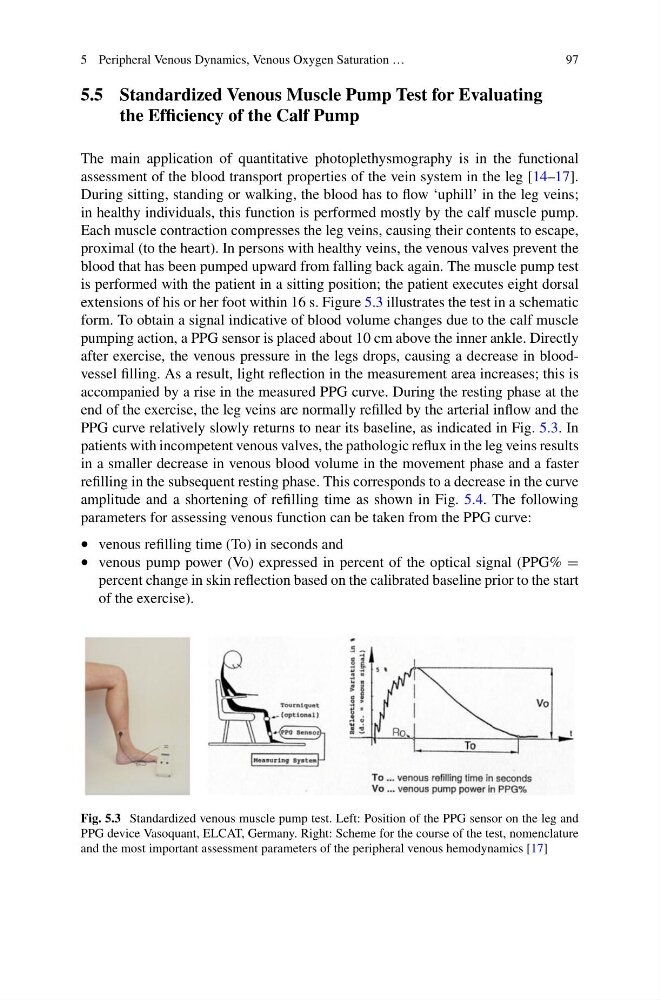
extensions of his or her foot within 16 s. Figure 5.3 illustrates the test in a schematic
form. To obtain a signal indicative of blood volume changes due to the calf muscle
pumping action, a PPG sensor is placed about 10 cm above the inner ankle. Directly
after exercise, the venous pressure in the legs drops, causing a decrease in bloodvessel filling. As a result, light reflection in the measurement area increases; this is
accompanied by a rise in the measured PPG curve. During the resting phase at the
end of the exercise, the leg veins are normally refilled by the arterial inflow and the
PPG curve relatively slowly returns to near its baseline, as indicated in Fig. 5.3. In
patients with incompetent venous valves, the pathologic reflux in the leg veins results
in a smaller decrease in venous blood volume in the movement phase and a faster
refilling in the subsequent resting phase. This corresponds to a decrease in the curve
amplitude and a shortening of refilling time as shown in Fig. 5.4. The following
parameters for assessing venous function can be taken from the PPG curve:
• venous refilling time (To) in seconds and
• venous pump power (Vo) expressed in percent of the optical signal (PPG% =
percent change in skin reflection based on the calibrated baseline prior to the start
of the exercise).
Fig. 5.3 Standardized venous muscle pump test. Left: Position of the PPG sensor on the leg and
PPG device Vasoquant, ELCAT, Germany. Right: Scheme for the course of the test, nomenclature
and the most important assessment parameters of the peripheral venous hemodynamics [17]
97
5.5 Standardized Venous Muscle Pump Test for Evaluating
the Efficiency of the Calf Pump
The main application of quantitative photoplethysmography is in the functional
assessment of the blood transport properties of the vein system in the leg [14–17].
During sitting, standing or walking, the blood has to flow ‘uphill’ in the leg veins;
in healthy individuals, this function is performed mostly by the calf muscle pump.
Each muscle contraction compresses the leg veins, causing their contents to escape,
proximal (to the heart). In persons with healthy veins, the venous valves prevent the
blood that has been pumped upward from falling back again. The muscle pump test
is performed with the patient in a sitting position; the patient executes eight dorsal
extensions of his or her foot within 16 s. Figure 5.3 illustrates the test in a schematic
form. To obtain a signal indicative of blood volume changes due to the calf muscle
pumping action, a PPG sensor is placed about 10 cm above the inner ankle. Directly
after exercise, the venous pressure in the legs drops, causing a decrease in bloodvessel filling. As a result, light reflection in the measurement area increases; this is
accompanied by a rise in the measured PPG curve. During the resting phase at the
end of the exercise, the leg veins are normally refilled by the arterial inflow and the
PPG curve relatively slowly returns to near its baseline, as indicated in Fig. 5.3. In
patients with incompetent venous valves, the pathologic reflux in the leg veins results
in a smaller decrease in venous blood volume in the movement phase and a faster
refilling in the subsequent resting phase. This corresponds to a decrease in the curve
amplitude and a shortening of refilling time as shown in Fig. 5.4. The following
parameters for assessing venous function can be taken from the PPG curve:
• venous refilling time (To) in seconds and
• venous pump power (Vo) expressed in percent of the optical signal (PPG% =
percent change in skin reflection based on the calibrated baseline prior to the start
of the exercise).
Fig. 5.3 Standardized venous muscle pump test. Left: Position of the PPG sensor on the leg and
PPG device Vasoquant, ELCAT, Germany. Right: Scheme for the course of the test, nomenclature
and the most important assessment parameters of the peripheral venous hemodynamics [17]