

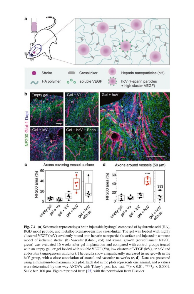
Fig. 7.4 (a) Schematic representing a brain injectable hydrogel composed of hyaluronic acid (HA),
RGD motif peptide, and metalloproteinase-sensitive cross-linker. The gel was loaded with highly
clustered VEGF (hcV) covalently bound onto heparin nanoparticle’s surface and injected in a mouse
model of ischemic stroke. (b) Vascular (Glut-1, red) and axonal growth (neurofilament NF200,
green) was evaluated 16 weeks after gel implantation and compared with control groups treated
with an empty gel, or gel loaded with soluble VEGF (Vs), low clusters of VEGF (lcV), or hcV and
endostatin (angiogenesis inhibitor). The results show a significantly increased tissue growth in the
hcV group, with a close association of axonal and vascular networks (c, d). Data are presented
using a minimum-to-maximum box plot. Each dot in the plots represents one animal, and p values
were determined by one-way ANOVA with Tukey’s post hoc test. **p < 0.01, ****p < 0.0001.
Scale bar, 100 μm. Figure reprinted from [25] with the permission from Elsevier
RGD motif peptide, and metalloproteinase-sensitive cross-linker. The gel was loaded with highly
clustered VEGF (hcV) covalently bound onto heparin nanoparticle’s surface and injected in a mouse
model of ischemic stroke. (b) Vascular (Glut-1, red) and axonal growth (neurofilament NF200,
green) was evaluated 16 weeks after gel implantation and compared with control groups treated
with an empty gel, or gel loaded with soluble VEGF (Vs), low clusters of VEGF (lcV), or hcV and
endostatin (angiogenesis inhibitor). The results show a significantly increased tissue growth in the
hcV group, with a close association of axonal and vascular networks (c, d). Data are presented
using a minimum-to-maximum box plot. Each dot in the plots represents one animal, and p values
were determined by one-way ANOVA with Tukey’s post hoc test. **p < 0.01, ****p < 0.0001.
Scale bar, 100 μm. Figure reprinted from [25] with the permission from Elsevier