

146
Cells
Culture conditions
Cardiac characterization
Vascular characterization
Ref.
hESC-CM hESC-CM
• 400,000 cells
• Percentage: 33–40%
Embryonic mouse fibroblasts
• 200,000–400,000 cells
• Percentage: 20–33%
hESC-EC or HUVECs
• 400,000 cells
• Percentage: 33–40%
• 50% PLLA: 50% PLGA size: 9 mm
3
(3 mm × 3 mm × 1 mm)
• Pore size: 212–600 μm
93% porosity
• Cultured for 2 weeks
• 8–10 μL culture media: Growth factor reduced
Matrigel mix (1:1)
• Simultaneously seeded
Advantages:
• Spontaneous beating occurred at day 4
• Ca
2+
impulse propagation in line with contraction
• Contractility is responsive to cardiac drugs like
isoproterenol and carbamylcholine
• Increased proliferation of cardiomyocytes in the
presence of endothelial cells
Limitations:
• Alignment of cardiomyocytes were not
characterized
• Electrophysiological properties not characterized
(i.e. conduction velocity and maximum captured
rate)
Advantages:
• No significant difference with between 1:1
and 1:2 (FB:EC)
• Tri-culture resulted in 5.12 lumen density
while endothelial was 1.57
• Implanted in vivo and becomes functional
with host vasculature
Limitations:
• Did not organize into blood vessels
without fibroblasts
• Mainly compact clusters
[13,
64]
hESC-CM
• 2,000,000 cells
• Percentage: 40%
Mouse embryonic fibroblasts
(MEF) or NHDF
• 2,000,000 cells
• Percentage: 40%
hESC-EC or HUVECs
• 1,000,000 cells
• Percentage: 20%
• Spheroid/cell clusters
• Cultured in low attachment plates
• Cultured for 3 days and 8 days
• Placed in vivo in a skeletal muscle defect and
nude rat hearts
Advantages:
• Human cardiac tissues patches contracted and
could be paced up to 2–3 Hz
• Increased collagen content in the tri-culture
• Increased force of contraction ~8 mN/mm
2
compared to 2 mN/mm
2
• Increased normalized force in the presence of all
three cell types
• Increased force in tri-culture with increased
differential strain
Limitations:
• Unorganized structure
• Lack of mechanical integrity
Advantages:
• Vessel structures increased in HuEB media
as compared to RPMI B27
• Increased CD31 staining compared to
cardiomyocyte only control
• Anastomosis with host vasculature in vivo
Limitations:
• Vessel length not assessed in vitro before
implantation
[111]
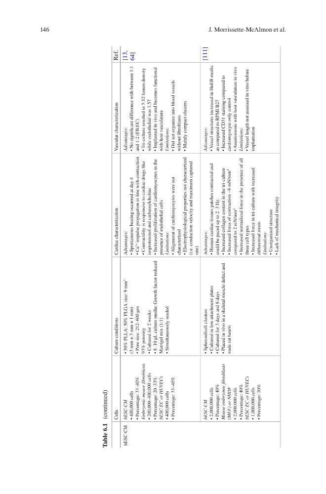
Table 6.1 (continued)
J. Morrissette-McAlmon et al.
Cells
Culture conditions
Cardiac characterization
Vascular characterization
Ref.
hESC-CM hESC-CM
• 400,000 cells
• Percentage: 33–40%
Embryonic mouse fibroblasts
• 200,000–400,000 cells
• Percentage: 20–33%
hESC-EC or HUVECs
• 400,000 cells
• Percentage: 33–40%
• 50% PLLA: 50% PLGA size: 9 mm
3
(3 mm × 3 mm × 1 mm)
• Pore size: 212–600 μm
93% porosity
• Cultured for 2 weeks
• 8–10 μL culture media: Growth factor reduced
Matrigel mix (1:1)
• Simultaneously seeded
Advantages:
• Spontaneous beating occurred at day 4
• Ca
2+
impulse propagation in line with contraction
• Contractility is responsive to cardiac drugs like
isoproterenol and carbamylcholine
• Increased proliferation of cardiomyocytes in the
presence of endothelial cells
Limitations:
• Alignment of cardiomyocytes were not
characterized
• Electrophysiological properties not characterized
(i.e. conduction velocity and maximum captured
rate)
Advantages:
• No significant difference with between 1:1
and 1:2 (FB:EC)
• Tri-culture resulted in 5.12 lumen density
while endothelial was 1.57
• Implanted in vivo and becomes functional
with host vasculature
Limitations:
• Did not organize into blood vessels
without fibroblasts
• Mainly compact clusters
[13,
64]
hESC-CM
• 2,000,000 cells
• Percentage: 40%
Mouse embryonic fibroblasts
(MEF) or NHDF
• 2,000,000 cells
• Percentage: 40%
hESC-EC or HUVECs
• 1,000,000 cells
• Percentage: 20%
• Spheroid/cell clusters
• Cultured in low attachment plates
• Cultured for 3 days and 8 days
• Placed in vivo in a skeletal muscle defect and
nude rat hearts
Advantages:
• Human cardiac tissues patches contracted and
could be paced up to 2–3 Hz
• Increased collagen content in the tri-culture
• Increased force of contraction ~8 mN/mm
2
compared to 2 mN/mm
2
• Increased normalized force in the presence of all
three cell types
• Increased force in tri-culture with increased
differential strain
Limitations:
• Unorganized structure
• Lack of mechanical integrity
Advantages:
• Vessel structures increased in HuEB media
as compared to RPMI B27
• Increased CD31 staining compared to
cardiomyocyte only control
• Anastomosis with host vasculature in vivo
Limitations:
• Vessel length not assessed in vitro before
implantation
[111]
Table 6.1 (continued)
J. Morrissette-McAlmon et al.