

145
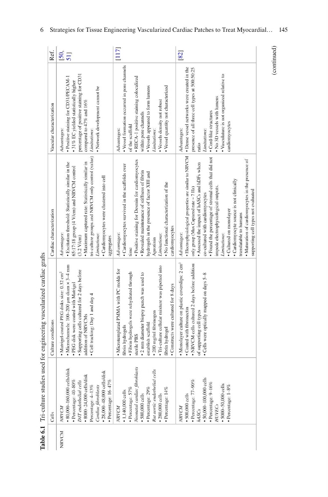
Table 6.1 Tri-culture studies used for engineering vascularized cardiac grafts
Cells
Culture conditions
Cardiac characterization
Vascular characterization
Ref.
NRVCM
NRVCM
• 80,000–160,000 cells/disk
• Percentage: 40–80%
D4T endothelial cells
• 8000–24,000 cells/disk
Percentage: 4–13%
Cardiac fibroblasts
• 28,000–48,000 cells/disk
• Percentage: 16–47%
• Matrigel-coated PEG disks size: 0.32 cm
2
• Microchannels: 100–200 μm diam × 3–4 mm
• PEG disk were coated with Matrigel
• Supporting cells cultured for 2 days before
addition of NRVCMs
• Cell tracking: Day 1 and day 4
Advantages:
• Excitation threshold: Statistically similar in the
65:17:18 group (4 V/cm) and NRVCM control
(3.2 V/cm)
• Maximum captured rate: Statistically similar in
tri-culture groups and NRVCM only control (value)
Limitations:
• Cardiomyocytes were clustered into cell
aggregates
Advantages:
• Positive staining for CD31/PECAM-1
• 31% EC yielded statistically higher
percentage of positive staining for CD31
compared to 47% and 16%
Limitations:
• Network development cannot be
[50,
51]
NRVCM
• 1,140,000 cells
• Percentage: 57%
Neonatal cardiac fibroblasts
• 580,000 cells
• Percentage: 29%
Rat aortic endothelial cells
• 280,000 cells
• Percentage: 14%
• Microtemplated PMMA with PC molds for
fibrin hydrogels
• Fibrin hydrogels were rehydrated through
sterile PBS
• 2 mm diameter biopsy punch was used to
establish scaffold
• 200 mg/ml fibrinogen
• Tri-culture cellular mixture was pipetted into
fibrin hydrogel
• Constructs were cultured for 8 days
Advantages:
• Cardiomyocytes survived in the scaffolds over
time
• Positive staining for Desmin for cardiomyocytes
• Revealed maintenance of stiffness of fibrin
hydrogels in the presence of factor XIII and
Aprotinin
Limitations:
• No functional characterization of the
cardiomyocytes
Advantages:
• Vessel formation occurred in pore channels
of the scaffold
• RECA-1 positive staining colocalized
within pore channels
• Vessels appeared to form lumens
Limitations:
• Vessels density not robust
• Vessel quantity not characterized
[117]
NRVCM
• 500,000 cells
• Percentage: 77–90%
hASCs
• 50,000–100,000 cells
• Percentage: 9–16%
HUVECs
• 5000–50,000 cells
• Percentage: 1–8%
• Monolayer culture on plastic coverslips: 2 cm
2
• Coated with fibronectin
• NRVCM cells cultured 2 days before addition
of supporting cell types
• Cells were optically mapped on days 5–8
Advantages:
• Electrophysiological properties are similar to NRVCM
only group (Max Captured rate ~ 7 Hz)
• Assessed the impact of hASCs and hDFs when
co-cultured with cardiomyocytes
• Found the percentage of stromal cells that did not
impede electrophysiological outputs.
Limitations:
• Cultured on monolayer
• Cardiomyocyte source is not clinically
translatable to humans
• Maturation of cardiomyocytes in the presence of
supporting cell types not evaluated
Advantages:
• Dense vessel networks were created in the
presence of all three cell types at 500:50:25
ratio
Limitations:
• Cord-like structures
• No 3D vessels with lumens
• Vasculature is not organized relative to
cardiomyocytes
[82]
(continued)
6 Strategies for Tissue Engineering Vascularized Cardiac Patches to Treat Myocardial…
Table 6.1 Tri-culture studies used for engineering vascularized cardiac grafts
Cells
Culture conditions
Cardiac characterization
Vascular characterization
Ref.
NRVCM
NRVCM
• 80,000–160,000 cells/disk
• Percentage: 40–80%
D4T endothelial cells
• 8000–24,000 cells/disk
Percentage: 4–13%
Cardiac fibroblasts
• 28,000–48,000 cells/disk
• Percentage: 16–47%
• Matrigel-coated PEG disks size: 0.32 cm
2
• Microchannels: 100–200 μm diam × 3–4 mm
• PEG disk were coated with Matrigel
• Supporting cells cultured for 2 days before
addition of NRVCMs
• Cell tracking: Day 1 and day 4
Advantages:
• Excitation threshold: Statistically similar in the
65:17:18 group (4 V/cm) and NRVCM control
(3.2 V/cm)
• Maximum captured rate: Statistically similar in
tri-culture groups and NRVCM only control (value)
Limitations:
• Cardiomyocytes were clustered into cell
aggregates
Advantages:
• Positive staining for CD31/PECAM-1
• 31% EC yielded statistically higher
percentage of positive staining for CD31
compared to 47% and 16%
Limitations:
• Network development cannot be
[50,
51]
NRVCM
• 1,140,000 cells
• Percentage: 57%
Neonatal cardiac fibroblasts
• 580,000 cells
• Percentage: 29%
Rat aortic endothelial cells
• 280,000 cells
• Percentage: 14%
• Microtemplated PMMA with PC molds for
fibrin hydrogels
• Fibrin hydrogels were rehydrated through
sterile PBS
• 2 mm diameter biopsy punch was used to
establish scaffold
• 200 mg/ml fibrinogen
• Tri-culture cellular mixture was pipetted into
fibrin hydrogel
• Constructs were cultured for 8 days
Advantages:
• Cardiomyocytes survived in the scaffolds over
time
• Positive staining for Desmin for cardiomyocytes
• Revealed maintenance of stiffness of fibrin
hydrogels in the presence of factor XIII and
Aprotinin
Limitations:
• No functional characterization of the
cardiomyocytes
Advantages:
• Vessel formation occurred in pore channels
of the scaffold
• RECA-1 positive staining colocalized
within pore channels
• Vessels appeared to form lumens
Limitations:
• Vessels density not robust
• Vessel quantity not characterized
[117]
NRVCM
• 500,000 cells
• Percentage: 77–90%
hASCs
• 50,000–100,000 cells
• Percentage: 9–16%
HUVECs
• 5000–50,000 cells
• Percentage: 1–8%
• Monolayer culture on plastic coverslips: 2 cm
2
• Coated with fibronectin
• NRVCM cells cultured 2 days before addition
of supporting cell types
• Cells were optically mapped on days 5–8
Advantages:
• Electrophysiological properties are similar to NRVCM
only group (Max Captured rate ~ 7 Hz)
• Assessed the impact of hASCs and hDFs when
co-cultured with cardiomyocytes
• Found the percentage of stromal cells that did not
impede electrophysiological outputs.
Limitations:
• Cultured on monolayer
• Cardiomyocyte source is not clinically
translatable to humans
• Maturation of cardiomyocytes in the presence of
supporting cell types not evaluated
Advantages:
• Dense vessel networks were created in the
presence of all three cell types at 500:50:25
ratio
Limitations:
• Cord-like structures
• No 3D vessels with lumens
• Vasculature is not organized relative to
cardiomyocytes
[82]
(continued)
6 Strategies for Tissue Engineering Vascularized Cardiac Patches to Treat Myocardial…