

as these dual-mode instruments become more available, the method will also be
more popular. Instruments with 3-condenser lens systems, which allow for flexible
control of semi-convergence angle and probe size, are increasingly available.
Complementary methods of “serial surface” imaging by SEM combined with
microtomy or FIB milling of plastic sections (covered in other chapters of this
book) are excellent techniques that have their own advantages including wide field
of view (FOV), lack of missing-wedge artifacts, etc. But the inherently lower
resolution of the SEM means that if it is desired to obtain all the detail that the stain
distribution provides, then STEM tomography is still the application of choice.
Because STEM is superior for identifying ultra-small gold labels and other
fiducial markers [54, 55], it also holds high promise for multiplex-immunolabeling
studies, where several sizes of fiducial or alternatively, fiducials of different metal
compositions, could be discriminated and identified.
For vitrified samples, CSTET is a brand new method, and we anticipate it will
meet a need for observing biological organelles within the context of their cellular
CET
CSTET
CSTET
CET
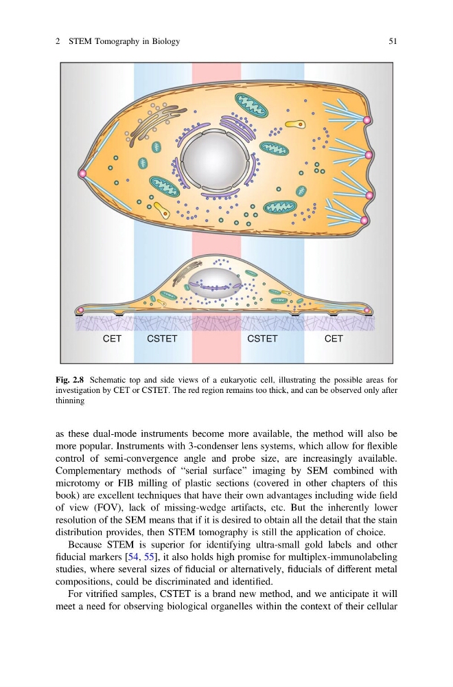
Fig. 2.8 Schematic top and side views of a eukaryotic cell, illustrating the possible areas for
investigation by CET or CSTET. The red region remains too thick, and can be observed only after
thinning
2 STEM Tomography in Biology
51
more popular. Instruments with 3-condenser lens systems, which allow for flexible
control of semi-convergence angle and probe size, are increasingly available.
Complementary methods of “serial surface” imaging by SEM combined with
microtomy or FIB milling of plastic sections (covered in other chapters of this
book) are excellent techniques that have their own advantages including wide field
of view (FOV), lack of missing-wedge artifacts, etc. But the inherently lower
resolution of the SEM means that if it is desired to obtain all the detail that the stain
distribution provides, then STEM tomography is still the application of choice.
Because STEM is superior for identifying ultra-small gold labels and other
fiducial markers [54, 55], it also holds high promise for multiplex-immunolabeling
studies, where several sizes of fiducial or alternatively, fiducials of different metal
compositions, could be discriminated and identified.
For vitrified samples, CSTET is a brand new method, and we anticipate it will
meet a need for observing biological organelles within the context of their cellular
CET
CSTET
CSTET
CET
Fig. 2.8 Schematic top and side views of a eukaryotic cell, illustrating the possible areas for
investigation by CET or CSTET. The red region remains too thick, and can be observed only after
thinning
2 STEM Tomography in Biology
51