

2.4 Conclusions and Future Prospects
It is recognized that STEM tomography holds enormous promise for thicker biological sections. Despite this, the method has not been widely applied. With the
development of computer controlled TEM/STEM instruments in the last two decades, high-performance STEM has became routine in a regular TEM column and
many more researchers now have the opportunity to apply STEM to biological
materials. At the Weizmann Institute, we have used exclusively STEM (rather than
TEM) for tomography of plastic sections (details of the experimental setup in
Sect. 2.5) since obtaining our Tecnai F20 TEM/STEM [95–99], and we believe that
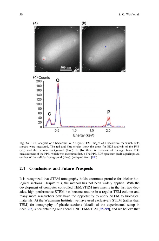
Fig. 2.7 EDS analysis of a bacterium. a, b Cryo-STEM images of a bacterium for which EDS
spectra were measured. The red and blue circles show the areas for EDS analysis of the PPB
(red) and the cellular background (blue). In (b), there is evidence of damage from EDS
measurement of the PPB, which was measured first. c The PPB EDS spectrum (red) superimposed
on that of the cellular background (blue). (Adapted from [84])
50
S. G. Wolf et al.
It is recognized that STEM tomography holds enormous promise for thicker biological sections. Despite this, the method has not been widely applied. With the
development of computer controlled TEM/STEM instruments in the last two decades, high-performance STEM has became routine in a regular TEM column and
many more researchers now have the opportunity to apply STEM to biological
materials. At the Weizmann Institute, we have used exclusively STEM (rather than
TEM) for tomography of plastic sections (details of the experimental setup in
Sect. 2.5) since obtaining our Tecnai F20 TEM/STEM [95–99], and we believe that
Fig. 2.7 EDS analysis of a bacterium. a, b Cryo-STEM images of a bacterium for which EDS
spectra were measured. The red and blue circles show the areas for EDS analysis of the PPB
(red) and the cellular background (blue). In (b), there is evidence of damage from EDS
measurement of the PPB, which was measured first. c The PPB EDS spectrum (red) superimposed
on that of the cellular background (blue). (Adapted from [84])
50
S. G. Wolf et al.