

(1) it requires a smaller r than 3D smoothing, and (2) it is possible to apply different
r values as a function of tilt angle, as suggested by [27].
In solving the inverse problem of reconstruction, extra free variables are required
to analytically account for irregularities in local grayscale value fluctuations as well
as missing data [2]. Thus, a recovery from ill-posedness (namely, regularization of
the inverse problem) is desirable for reducing reconstruction artefact. The discrete
algebraic reconstruction technique (DART) approaches the problem by drastically
reducing the possible range of intensity values in the whole reconstructed image [2,
3]. Although this approach effectively reduces artefacts, it drastically contracts the
intensity grayscale values of the reconstructed volume to a small set of discrete
values, which may cause problems in the interpretation of complex biological
structures. Compressive sensing approaches to the regularization of the inverse
problem of reconstruction have recently been described. Reference [17] presented a
reconstruction algorithm that uses total variation minimization (TVM) regularization
for reducing the overall range of intensity values. This method can produce a
reconstructed volume with a larger set of discrete grayscale values than DART.
However, the grayscale value impairment affects the whole image, which is not
desirable when only selected objects in the tomogram are causing the artefacts.
Pre-reconstruction Nonlinear Anisotropic diffusion (pre-NAD) may be used for
contrast enhancement. For pre-NAD [27], the magnitude of r determines the spatial
interval over which the structure tensor is calculated [1, 25, 33]. Small values of r
cause negligible smoothing of the image. Large values of r reduce noise but may
obscure significant features, such as edges and discontinuities. Pre-NAD compensates
for the variable projected depth through the sample by using a specific smoothing
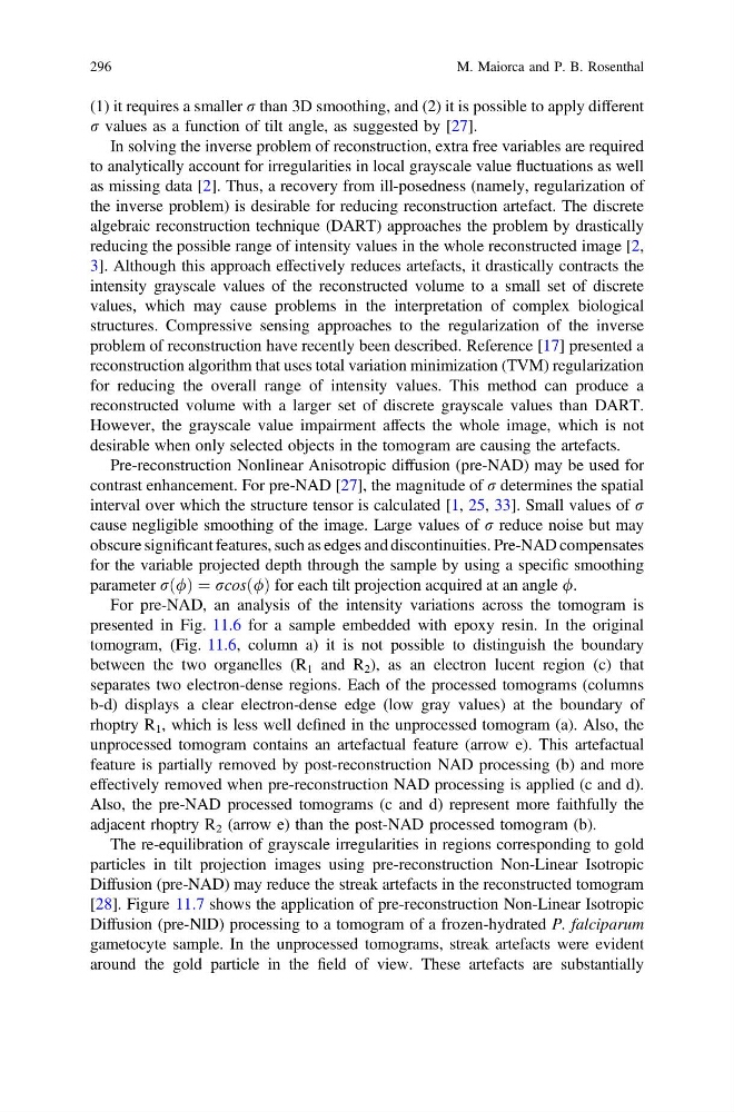
parameter rð/Þ ¼ rcosð/Þ for each tilt projection acquired at an angle /.
For pre-NAD, an analysis of the intensity variations across the tomogram is
presented in Fig. 11.6 for a sample embedded with epoxy resin. In the original
tomogram, (Fig. 11.6, column a) it is not possible to distinguish the boundary
between the two organelles (R 1 and R 2 ), as an electron lucent region (c) that
separates two electron-dense regions. Each of the processed tomograms (columns
b-d) displays a clear electron-dense edge (low gray values) at the boundary of
rhoptry R 1 , which is less well defined in the unprocessed tomogram (a). Also, the
unprocessed tomogram contains an artefactual feature (arrow e). This artefactual
feature is partially removed by post-reconstruction NAD processing (b) and more
effectively removed when pre-reconstruction NAD processing is applied (c and d).
Also, the pre-NAD processed tomograms (c and d) represent more faithfully the
adjacent rhoptry R 2 (arrow e) than the post-NAD processed tomogram (b).
The re-equilibration of grayscale irregularities in regions corresponding to gold
particles in tilt projection images using pre-reconstruction Non-Linear Isotropic
Diffusion (pre-NAD) may reduce the streak artefacts in the reconstructed tomogram
[28]. Figure 11.7 shows the application of pre-reconstruction Non-Linear Isotropic
Diffusion (pre-NID) processing to a tomogram of a frozen-hydrated P. falciparum
gametocyte sample. In the unprocessed tomograms, streak artefacts were evident
around the gold particle in the field of view. These artefacts are substantially
296
M. Maiorca and P. B. Rosenthal
r values as a function of tilt angle, as suggested by [27].
In solving the inverse problem of reconstruction, extra free variables are required
to analytically account for irregularities in local grayscale value fluctuations as well
as missing data [2]. Thus, a recovery from ill-posedness (namely, regularization of
the inverse problem) is desirable for reducing reconstruction artefact. The discrete
algebraic reconstruction technique (DART) approaches the problem by drastically
reducing the possible range of intensity values in the whole reconstructed image [2,
3]. Although this approach effectively reduces artefacts, it drastically contracts the
intensity grayscale values of the reconstructed volume to a small set of discrete
values, which may cause problems in the interpretation of complex biological
structures. Compressive sensing approaches to the regularization of the inverse
problem of reconstruction have recently been described. Reference [17] presented a
reconstruction algorithm that uses total variation minimization (TVM) regularization
for reducing the overall range of intensity values. This method can produce a
reconstructed volume with a larger set of discrete grayscale values than DART.
However, the grayscale value impairment affects the whole image, which is not
desirable when only selected objects in the tomogram are causing the artefacts.
Pre-reconstruction Nonlinear Anisotropic diffusion (pre-NAD) may be used for
contrast enhancement. For pre-NAD [27], the magnitude of r determines the spatial
interval over which the structure tensor is calculated [1, 25, 33]. Small values of r
cause negligible smoothing of the image. Large values of r reduce noise but may
obscure significant features, such as edges and discontinuities. Pre-NAD compensates
for the variable projected depth through the sample by using a specific smoothing
parameter rð/Þ ¼ rcosð/Þ for each tilt projection acquired at an angle /.
For pre-NAD, an analysis of the intensity variations across the tomogram is
presented in Fig. 11.6 for a sample embedded with epoxy resin. In the original
tomogram, (Fig. 11.6, column a) it is not possible to distinguish the boundary
between the two organelles (R 1 and R 2 ), as an electron lucent region (c) that
separates two electron-dense regions. Each of the processed tomograms (columns
b-d) displays a clear electron-dense edge (low gray values) at the boundary of
rhoptry R 1 , which is less well defined in the unprocessed tomogram (a). Also, the
unprocessed tomogram contains an artefactual feature (arrow e). This artefactual
feature is partially removed by post-reconstruction NAD processing (b) and more
effectively removed when pre-reconstruction NAD processing is applied (c and d).
Also, the pre-NAD processed tomograms (c and d) represent more faithfully the
adjacent rhoptry R 2 (arrow e) than the post-NAD processed tomogram (b).
The re-equilibration of grayscale irregularities in regions corresponding to gold
particles in tilt projection images using pre-reconstruction Non-Linear Isotropic
Diffusion (pre-NAD) may reduce the streak artefacts in the reconstructed tomogram
[28]. Figure 11.7 shows the application of pre-reconstruction Non-Linear Isotropic
Diffusion (pre-NID) processing to a tomogram of a frozen-hydrated P. falciparum
gametocyte sample. In the unprocessed tomograms, streak artefacts were evident
around the gold particle in the field of view. These artefacts are substantially
296
M. Maiorca and P. B. Rosenthal