

(Fig. 11.4b and detail b’) or Gaussian filtered (Fig. 11.4c and detail c’). Both the EED
and Gaussian filtered images show an increased contrast. However, EED filtering
better preserves small details blurred out by Gaussian filtering, for example glycoproteins (green arrows) and the inner matrix layer (red arrows).
11.3.5 Bilateral Filter
The bilateral filter was first introduced by [42], aiming to combine Gaussian diffusion with the elimination of noise-related small pixel intensity variations. The
bilateral filter was adapted for electron tomography by [24] and implemented in
EMAN2 [41].
In practice, the intensity value at each pixel in an image is replaced by a
Gaussian-weighted average of intensity values from pixels within a neighbourhood
n. The weights depend on Euclidean distance of pixels, and on the intensity value
differences. Using notation from [24],
I out ð r
! Þ ¼
Z
exp À
j n
! À r
! j
2
2r 2
1
(
)
exp À
jIð n
!
Þ À Ið r
! Þj
2
2r 2
2
(
)
 Ið n
!
Þd n
! ð11:13Þ
Ið r
! Þ is the input image, expðj n
! À r
! j
2 Þ=ð2r
2
1 Þ is the Euclidean distance contribution, and expðjIð n
!
Þ À Ið r
! Þj
2 Þ=ð2r
2
2 Þ is the intensity value difference.
Furthermore, variations of the bilateral filter can include an edge detection paradigm
[35]
The parameter r 1 controls the extent of the Gaussian diffusion, and a larger r 1
causes severe smoothing. r 2 controls the discrimination of intensity value differences. Larger pixel intensity value variations are mainly from true features and
smaller pixel intensity value variations are contributed by noise. This filter has the
property of removing isolated pixels, without the need of aggressive Gaussian
diffusion.
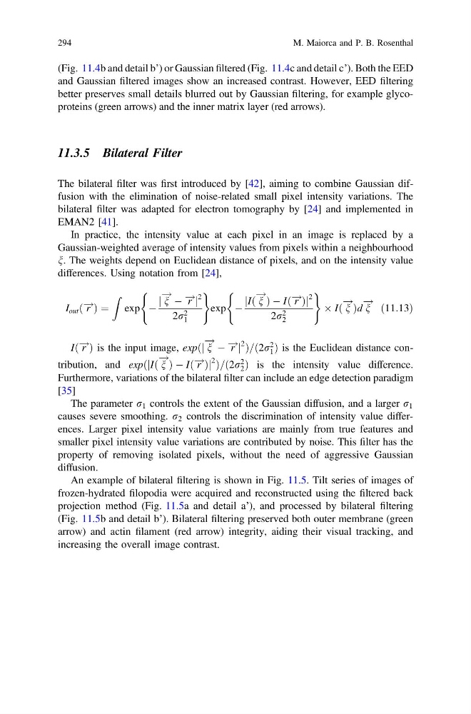
An example of bilateral filtering is shown in Fig. 11.5. Tilt series of images of
frozen-hydrated filopodia were acquired and reconstructed using the filtered back
projection method (Fig. 11.5a and detail a’), and processed by bilateral filtering
(Fig. 11.5b and detail b’). Bilateral filtering preserved both outer membrane (green
arrow) and actin filament (red arrow) integrity, aiding their visual tracking, and
increasing the overall image contrast.
294
M. Maiorca and P. B. Rosenthal
and Gaussian filtered images show an increased contrast. However, EED filtering
better preserves small details blurred out by Gaussian filtering, for example glycoproteins (green arrows) and the inner matrix layer (red arrows).
11.3.5 Bilateral Filter
The bilateral filter was first introduced by [42], aiming to combine Gaussian diffusion with the elimination of noise-related small pixel intensity variations. The
bilateral filter was adapted for electron tomography by [24] and implemented in
EMAN2 [41].
In practice, the intensity value at each pixel in an image is replaced by a
Gaussian-weighted average of intensity values from pixels within a neighbourhood
n. The weights depend on Euclidean distance of pixels, and on the intensity value
differences. Using notation from [24],
I out ð r
! Þ ¼
Z
exp À
j n
! À r
! j
2
2r 2
1
(
)
exp À
jIð n
!
Þ À Ið r
! Þj
2
2r 2
2
(
)
 Ið n
!
Þd n
! ð11:13Þ
Ið r
! Þ is the input image, expðj n
! À r
! j
2 Þ=ð2r
2
1 Þ is the Euclidean distance contribution, and expðjIð n
!
Þ À Ið r
! Þj
2 Þ=ð2r
2
2 Þ is the intensity value difference.
Furthermore, variations of the bilateral filter can include an edge detection paradigm
[35]
The parameter r 1 controls the extent of the Gaussian diffusion, and a larger r 1
causes severe smoothing. r 2 controls the discrimination of intensity value differences. Larger pixel intensity value variations are mainly from true features and
smaller pixel intensity value variations are contributed by noise. This filter has the
property of removing isolated pixels, without the need of aggressive Gaussian
diffusion.
An example of bilateral filtering is shown in Fig. 11.5. Tilt series of images of
frozen-hydrated filopodia were acquired and reconstructed using the filtered back
projection method (Fig. 11.5a and detail a’), and processed by bilateral filtering
(Fig. 11.5b and detail b’). Bilateral filtering preserved both outer membrane (green
arrow) and actin filament (red arrow) integrity, aiding their visual tracking, and
increasing the overall image contrast.
294
M. Maiorca and P. B. Rosenthal