

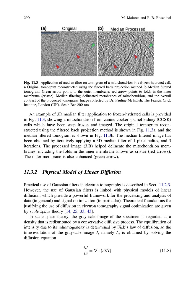
An example of 3D median filter application to frozen-hydrated cells is provided
in Fig. 11.3, showing a mitochondrion from canine cocker spaniel kidney (CCSK)
cells which have been snap frozen and imaged. The original tomogram reconstructed using the filtered back projection method is shown in Fig. 11.3a, and the
median filtered tomogram is shown in Fig. 11.3b. The median filtered image has
been obtained by iteratively applying a 3D median filter of 1 pixel radius, and 3
iterations. The processed image (3.B) helped delineate the mitochondrion membranes, including the folds in the inner membrane known as cristae (red arrows).
The outer membrane is also enhanced (green arrow).
11.3.2 Physical Model of Linear Diffusion
Practical use of Gaussian filters in electron tomography is described in Sect. 11.2.3.
However, the use of Gaussian filters is linked with physical models of linear
diffusion, which provide a powerful framework for the processing and analysis of
data (in general) and signal optimization (in particular). Theoretical foundations for
justifying the use of diffusion in electron tomography signal optimization are given
by scale space theory [14, 25, 33, 43].
In scale space theory, the grayscale image of the specimen is regarded as a
density that is redistributed by a conservative diffusive process. The equilibration of
intensity due to its inhomogeneity is determined by Fick’s law of diffusion, so the
time-evolution of the grayscale image I, namely I t , is obtained by solving the
diffusion equation
@I
@t
¼ r Á ðcrIÞ
ð 11:8Þ
Fig. 11.3 Application of median filter on tomogram of a mitochondrion in a frozen-hydrated cell.
a Original tomogram reconstructed using the filtered back projection method. b Median filtered
tomogram. Green arrow points to the outer membrane; red arrow points to folds in the inner
membrane (cristae). Median filtering delineated membranes of mitochondrion, and the overall
contrast of the processed tomogram. Image collected by Dr. Pauline McIntosh, The Francis Crick
Institute, London (UK). Scale Bar 200 nm
290
M. Maiorca and P. B. Rosenthal
in Fig. 11.3, showing a mitochondrion from canine cocker spaniel kidney (CCSK)
cells which have been snap frozen and imaged. The original tomogram reconstructed using the filtered back projection method is shown in Fig. 11.3a, and the
median filtered tomogram is shown in Fig. 11.3b. The median filtered image has
been obtained by iteratively applying a 3D median filter of 1 pixel radius, and 3
iterations. The processed image (3.B) helped delineate the mitochondrion membranes, including the folds in the inner membrane known as cristae (red arrows).
The outer membrane is also enhanced (green arrow).
11.3.2 Physical Model of Linear Diffusion
Practical use of Gaussian filters in electron tomography is described in Sect. 11.2.3.
However, the use of Gaussian filters is linked with physical models of linear
diffusion, which provide a powerful framework for the processing and analysis of
data (in general) and signal optimization (in particular). Theoretical foundations for
justifying the use of diffusion in electron tomography signal optimization are given
by scale space theory [14, 25, 33, 43].
In scale space theory, the grayscale image of the specimen is regarded as a
density that is redistributed by a conservative diffusive process. The equilibration of
intensity due to its inhomogeneity is determined by Fick’s law of diffusion, so the
time-evolution of the grayscale image I, namely I t , is obtained by solving the
diffusion equation
@I
@t
¼ r Á ðcrIÞ
ð 11:8Þ
Fig. 11.3 Application of median filter on tomogram of a mitochondrion in a frozen-hydrated cell.
a Original tomogram reconstructed using the filtered back projection method. b Median filtered
tomogram. Green arrow points to the outer membrane; red arrow points to folds in the inner
membrane (cristae). Median filtering delineated membranes of mitochondrion, and the overall
contrast of the processed tomogram. Image collected by Dr. Pauline McIntosh, The Francis Crick
Institute, London (UK). Scale Bar 200 nm
290
M. Maiorca and P. B. Rosenthal