

axonal projections [58]. Despite the great variety of inhibitory interneurons in cortex,
almost all of the inhibitory myelinated axons were found to belong to one interneuron
type, the parvalbumin basket cells. In addition, immunofluorescent AT analysis
shows that the myelin on these interneurons has a different organization (e.g. shorter
nodes of Ranvier and internodes) and protein composition (higher MBP content)
compared to excitatory axons. These findings challenge the notion that cortical
myelin is mostly restricted to long-range excitatory axons in cortex and highlight the
need to further explore the functional consequences of interneuron myelination.
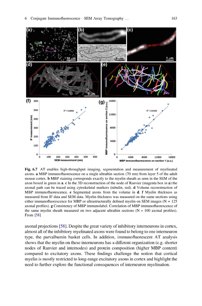
Fig. 6.7 AT enables high-throughput imaging, segmentation and measurement of myelinated
axons. a MBP immunofluorescence on a single ultrathin section (70 nm) from layer 5 of the adult
mouse cortex. b MBP staining corresponds exactly to the myelin sheath as seen in the SEM of the
axon boxed in green in a. c In the 3D reconstruction of the node of Ranvier (magenta box in a) the
axonal path can be traced using cytoskeletal markers (tubulin, red). d Volume reconstruction of
MBP immunofluorescence. e Segmented axons from the volume in d. f Myelin thickness as
measured from IF data and SEM data. Myelin thickness was measured on the same sections using
either immunofluorescence for MBP or ultrastructurally defined myelin on SEM images (N = 125
axonal profiles). g Consistency of MBP immunolabel. Correlation of MBP immunofluorescence of
the same myelin sheath measured on two adjacent ultrathin sections (N = 100 axonal profiles).
From [58]
6 Conjugate Immunofluorescence—SEM Array Tomography …
163
almost all of the inhibitory myelinated axons were found to belong to one interneuron
type, the parvalbumin basket cells. In addition, immunofluorescent AT analysis
shows that the myelin on these interneurons has a different organization (e.g. shorter
nodes of Ranvier and internodes) and protein composition (higher MBP content)
compared to excitatory axons. These findings challenge the notion that cortical
myelin is mostly restricted to long-range excitatory axons in cortex and highlight the
need to further explore the functional consequences of interneuron myelination.
Fig. 6.7 AT enables high-throughput imaging, segmentation and measurement of myelinated
axons. a MBP immunofluorescence on a single ultrathin section (70 nm) from layer 5 of the adult
mouse cortex. b MBP staining corresponds exactly to the myelin sheath as seen in the SEM of the
axon boxed in green in a. c In the 3D reconstruction of the node of Ranvier (magenta box in a) the
axonal path can be traced using cytoskeletal markers (tubulin, red). d Volume reconstruction of
MBP immunofluorescence. e Segmented axons from the volume in d. f Myelin thickness as
measured from IF data and SEM data. Myelin thickness was measured on the same sections using
either immunofluorescence for MBP or ultrastructurally defined myelin on SEM images (N = 125
axonal profiles). g Consistency of MBP immunolabel. Correlation of MBP immunofluorescence of
the same myelin sheath measured on two adjacent ultrathin sections (N = 100 axonal profiles).
From [58]
6 Conjugate Immunofluorescence—SEM Array Tomography …
163