

6.7 Variations of the Method
Conjugate IF-SEM array tomography on freeze-substituted and Lowicryl embedded
tissue enables the use of multiple antibodies against a variety of targets that can be
applied, for example, to identify and distinguish between excitatory and inhibitory
mammalian synapses, as well as to further explore synaptic diversity within these
broad categories. However, specific research questions that require only a small
subset of antibodies, may be more efficiently addressed using other tissue preparations. For example, as noted above, the antigenicity of GABA and other small
molecules is well preserved in tissue prepared using standard electron microscopy
procedures, including osmium postfixation and embedding in Epon. In fact, there is
vast literature that has used GABA immunostaining and TEM to study the development, distribution and plasticity of the inhibitory GABA circuitry in a variety of
animal species (for example, [61–64]). Therefore, when the focus of study is the
brain GABA network, or another target with similarly preserved antigenicity (e.g.
neuropeptides, [24]), conjugate IF-SEM array tomography can be performed on
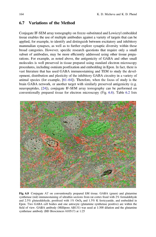
conventionally prepared tissue for electron microscopy (Fig. 6.8). Table 6.2 lists
Fig. 6.8 Conjugate AT on conventionally prepared EM tissue. GABA (green) and glutamine
synthetase (red) immunostaining of ultrathin sections from rat cortex fixed with 2% formaldehyde
and 2.5% glutaraldehyde, postfixed with 1% OsO 4 and 1.5% K ferricyanide, and embedded in
Epon. Two GABA cell bodies and one astrocyte (glutamine synthetase positive) are within the
field of view. GABA antibody (Millipore AB131) was used at 1:300 dilution and the glutamine
synthetase antibody (BD Biosciences 610517) at 1:25
164
K. D. Micheva and K. D. Phend
Conjugate IF-SEM array tomography on freeze-substituted and Lowicryl embedded
tissue enables the use of multiple antibodies against a variety of targets that can be
applied, for example, to identify and distinguish between excitatory and inhibitory
mammalian synapses, as well as to further explore synaptic diversity within these
broad categories. However, specific research questions that require only a small
subset of antibodies, may be more efficiently addressed using other tissue preparations. For example, as noted above, the antigenicity of GABA and other small
molecules is well preserved in tissue prepared using standard electron microscopy
procedures, including osmium postfixation and embedding in Epon. In fact, there is
vast literature that has used GABA immunostaining and TEM to study the development, distribution and plasticity of the inhibitory GABA circuitry in a variety of
animal species (for example, [61–64]). Therefore, when the focus of study is the
brain GABA network, or another target with similarly preserved antigenicity (e.g.
neuropeptides, [24]), conjugate IF-SEM array tomography can be performed on
conventionally prepared tissue for electron microscopy (Fig. 6.8). Table 6.2 lists
Fig. 6.8 Conjugate AT on conventionally prepared EM tissue. GABA (green) and glutamine
synthetase (red) immunostaining of ultrathin sections from rat cortex fixed with 2% formaldehyde
and 2.5% glutaraldehyde, postfixed with 1% OsO 4 and 1.5% K ferricyanide, and embedded in
Epon. Two GABA cell bodies and one astrocyte (glutamine synthetase positive) are within the
field of view. GABA antibody (Millipore AB131) was used at 1:300 dilution and the glutamine
synthetase antibody (BD Biosciences 610517) at 1:25
164
K. D. Micheva and K. D. Phend