

correct allocation of immunofluorescence to ultrastructural tissue compartments.
Ultrathin sectioning also ensures that all sections from a tissue volume are stained
and imaged under identical conditions, thus eliminating possible depth-related bias
in working with thicker samples.
Another advantage of physical sectioning is that the use of such ultrathin sections facilitates the elution of antibodies off the section array, which enables multiple applications of different antibodies to the same serial sections. This greatly
increases the number of proteins that can be visualized in one sample (more than 30
different antibodies per sample have been imaged by us) and therefore substantially
expands the scope of research questions that can be addressed.
In array tomography, the serial sections are mounted on a hard substrate
(coverslips) as opposed to grids. This allows the relatively easy collection of long
uninterrupted series (hundreds to thousands of sections), because coverslips provide
a much larger area and also a better support for the sections. Number 1.5 coverslips
(0.17 mm thickness) provide the ideal substrate for high-performance microscope
objectives and are preferred for section collection for array tomography. And while
the section arrays on coverslips cannot be imaged in a TEM, recent technological
advances enable excellent resolution with a field emission scanning electron
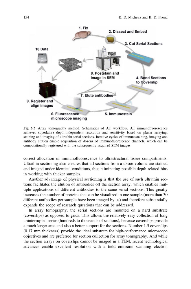
Fig. 6.3 Array tomography method. Schematics of AT workflow. AT immunofluorescence
achieves superlative depth-independent resolution and sensitivity based on planar arraying,
staining and imaging of ultrathin serial sections. Iterative cycles of immunostaining, imaging and
antibody elution enable acquisition of dozens of immunofluorescence channels, which can be
computationally registered with the subsequently acquired SEM images
154
K. D. Micheva and K. D. Phend
Ultrathin sectioning also ensures that all sections from a tissue volume are stained
and imaged under identical conditions, thus eliminating possible depth-related bias
in working with thicker samples.
Another advantage of physical sectioning is that the use of such ultrathin sections facilitates the elution of antibodies off the section array, which enables multiple applications of different antibodies to the same serial sections. This greatly
increases the number of proteins that can be visualized in one sample (more than 30
different antibodies per sample have been imaged by us) and therefore substantially
expands the scope of research questions that can be addressed.
In array tomography, the serial sections are mounted on a hard substrate
(coverslips) as opposed to grids. This allows the relatively easy collection of long
uninterrupted series (hundreds to thousands of sections), because coverslips provide
a much larger area and also a better support for the sections. Number 1.5 coverslips
(0.17 mm thickness) provide the ideal substrate for high-performance microscope
objectives and are preferred for section collection for array tomography. And while
the section arrays on coverslips cannot be imaged in a TEM, recent technological
advances enable excellent resolution with a field emission scanning electron
Fig. 6.3 Array tomography method. Schematics of AT workflow. AT immunofluorescence
achieves superlative depth-independent resolution and sensitivity based on planar arraying,
staining and imaging of ultrathin serial sections. Iterative cycles of immunostaining, imaging and
antibody elution enable acquisition of dozens of immunofluorescence channels, which can be
computationally registered with the subsequently acquired SEM images
154
K. D. Micheva and K. D. Phend