

6.3 Array Tomography: A Different Kind of Tomography
‘Tomography’ comes from the Greek word tomos, meaning a cut, slice or section,
and refers to methods used for reconstructing the three-dimensional structure of an
object from two-dimensional series of images. With electron tomography the slicing
is achieved by tilting the stage and imaging the sample at different angles; with array
tomography, the slicing is physical and involves actual cutting of ultrathin sections
(Fig. 6.3). Both electron tomography and array tomography require sophisticated
computational approaches for volume reconstruction.
The ultrathin physical sectioning of the sample used in AT offers a number of
advantages. Of primary importance are the high resolution and depth independence
of the immunofluorescence imaging (Fig. 6.4). The largest improvement in resolution comes from the physical section thickness (usually 70 nm), which is about 10
times thinner than the effective z-axis resolution of a confocal microscope [37]. The
lateral resolution is also improved (*200 nm) due to reduced light scatter in
ultrathin sections, as well as the fact that imaging occurs at the design conditions for
the high numerical-aperture objective used (i.e. the immediate contact between
specimen and coverslip). Lateral resolution can be further improved to about
100 nm either by deconvolution [11], or by the use of structured illumination
microscopy (SIM; [38, 39]; Fig. 6.4b). The high resolution of fluorescence imaging
facilitates registration of the light and electron microscopic images and ensures the
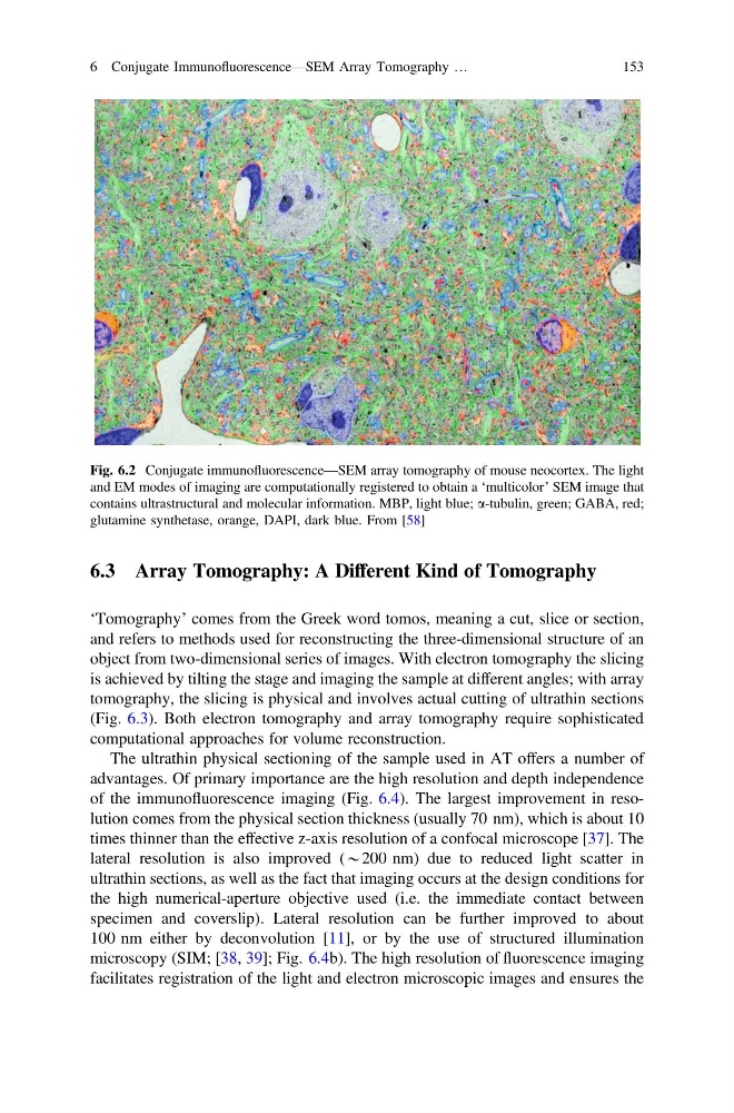
Fig. 6.2 Conjugate immunofluorescence—SEM array tomography of mouse neocortex. The light
and EM modes of imaging are computationally registered to obtain a ‘multicolor’ SEM image that
contains ultrastructural and molecular information. MBP, light blue; a-tubulin, green; GABA, red;
glutamine synthetase, orange, DAPI, dark blue. From [58]
6 Conjugate Immunofluorescence—SEM Array Tomography …
153
‘Tomography’ comes from the Greek word tomos, meaning a cut, slice or section,
and refers to methods used for reconstructing the three-dimensional structure of an
object from two-dimensional series of images. With electron tomography the slicing
is achieved by tilting the stage and imaging the sample at different angles; with array
tomography, the slicing is physical and involves actual cutting of ultrathin sections
(Fig. 6.3). Both electron tomography and array tomography require sophisticated
computational approaches for volume reconstruction.
The ultrathin physical sectioning of the sample used in AT offers a number of
advantages. Of primary importance are the high resolution and depth independence
of the immunofluorescence imaging (Fig. 6.4). The largest improvement in resolution comes from the physical section thickness (usually 70 nm), which is about 10
times thinner than the effective z-axis resolution of a confocal microscope [37]. The
lateral resolution is also improved (*200 nm) due to reduced light scatter in
ultrathin sections, as well as the fact that imaging occurs at the design conditions for
the high numerical-aperture objective used (i.e. the immediate contact between
specimen and coverslip). Lateral resolution can be further improved to about
100 nm either by deconvolution [11], or by the use of structured illumination
microscopy (SIM; [38, 39]; Fig. 6.4b). The high resolution of fluorescence imaging
facilitates registration of the light and electron microscopic images and ensures the
Fig. 6.2 Conjugate immunofluorescence—SEM array tomography of mouse neocortex. The light
and EM modes of imaging are computationally registered to obtain a ‘multicolor’ SEM image that
contains ultrastructural and molecular information. MBP, light blue; a-tubulin, green; GABA, red;
glutamine synthetase, orange, DAPI, dark blue. From [58]
6 Conjugate Immunofluorescence—SEM Array Tomography …
153