

longer needed to stabilize the lipids, and can be omitted, thus greatly improving antigenicity without a parallel decline in ultrastructural preservation. To fully benefit from
the low temperature dehydration, samples need also be infiltrated with resin and
polymerized at low temperature, which is not possible with the conventional epoxy
resins. Instead, other resins, such as the acrylic Lowicryl resins, can be used as they
remain liquid and can polymerize at low temperatures. These so-called
“freeze-substitution” methods have allowed the immmunodetection of a variety of
antibodies with nanometer precision, using immunogold EM (for example, [31–36]).
Because of the well preserved antigenicity and ultrastructure, freeze-substitution followed by embedding in Lowicryl produces tissue samples that are suitable not only for
electron microscopy, but also for immunofluorescence, as recently achieved with the
method of array tomography [18]. Conjugate array tomography, as this specific
application is named, uses Lowicryl HM20 to ensure good preservation of tissue
ultrastructure and antigenicity and thus enable imaging of both the molecular content
and the ultrastructure of tissues (Fig. 6.2). This method is based on physical ultrathin
serial sectioning, immunostaining and acquiring fluorescence and electron microscopy
images of resin embedded tissues, followed by computational volume reconstruction
and analysis.
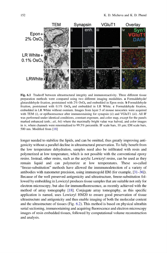
Fig. 6.1 Tradeoff between ultrastructural integrity and immunoreactivity. Three different tissue
preparation methods were compared using two different imaging modalities. a Formaldehyde/
glutaraldehyde fixation, poststained with 2% OsO 4 and embedded in Epon resin. b Formaldehyde
fixation, poststained with 0.1% OsO 4 and embedded in LR White. c Formaldehyde fixation,
embedded in LR White without osmium. Images from layer 5 of mouse neocortex were acquired
with TEM (i), or epifluorescence after immunostaining for synapsin (ii) and VGluT1 (iii). All IF
was performed under identical conditions, constant exposure, and color map, except for the panels
marked enhanced (enh.; aii, bii) where the maximally bright value was halved, and color images
in iv, where channels were renormalized to 99.5% percentile. IF scale bars, 10 lm; EM scale bars,
500 nm. Modified from [18]
152
K. D. Micheva and K. D. Phend
the low temperature dehydration, samples need also be infiltrated with resin and
polymerized at low temperature, which is not possible with the conventional epoxy
resins. Instead, other resins, such as the acrylic Lowicryl resins, can be used as they
remain liquid and can polymerize at low temperatures. These so-called
“freeze-substitution” methods have allowed the immmunodetection of a variety of
antibodies with nanometer precision, using immunogold EM (for example, [31–36]).
Because of the well preserved antigenicity and ultrastructure, freeze-substitution followed by embedding in Lowicryl produces tissue samples that are suitable not only for
electron microscopy, but also for immunofluorescence, as recently achieved with the
method of array tomography [18]. Conjugate array tomography, as this specific
application is named, uses Lowicryl HM20 to ensure good preservation of tissue
ultrastructure and antigenicity and thus enable imaging of both the molecular content
and the ultrastructure of tissues (Fig. 6.2). This method is based on physical ultrathin
serial sectioning, immunostaining and acquiring fluorescence and electron microscopy
images of resin embedded tissues, followed by computational volume reconstruction
and analysis.
Fig. 6.1 Tradeoff between ultrastructural integrity and immunoreactivity. Three different tissue
preparation methods were compared using two different imaging modalities. a Formaldehyde/
glutaraldehyde fixation, poststained with 2% OsO 4 and embedded in Epon resin. b Formaldehyde
fixation, poststained with 0.1% OsO 4 and embedded in LR White. c Formaldehyde fixation,
embedded in LR White without osmium. Images from layer 5 of mouse neocortex were acquired
with TEM (i), or epifluorescence after immunostaining for synapsin (ii) and VGluT1 (iii). All IF
was performed under identical conditions, constant exposure, and color map, except for the panels
marked enhanced (enh.; aii, bii) where the maximally bright value was halved, and color images
in iv, where channels were renormalized to 99.5% percentile. IF scale bars, 10 lm; EM scale bars,
500 nm. Modified from [18]
152
K. D. Micheva and K. D. Phend