

intersystem crossings (S 1 ~~> T 1 as well as T 1 ! S 0 ) through heavy atom effect
[46] as evidenced by the fast fluorescence decay of excited pyrene in Py•Xe@OA 2
compared to the same in absence of Xe (Py@OA 2 ). Computational modeling and
NMR studies supported the postulate that Py and at least one Xe atom were present
within the OA capsule (Fig. 10), which resulted in high degree of RTP. Py lacked
any significant RTP under all other conditions wherein Xe was not present: as a free
luminophore, complexed to OA in aerated and N 2 -purged solutions (Fig. 10, black,
blue, and green).
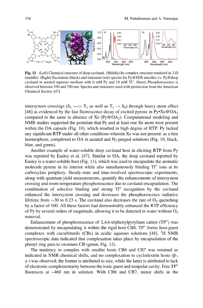
Another example of water-soluble deep cavitand host in eliciting RTP from Py
was reported by Easley et al. [47]. Similar to OA, the deep cavitand reported by
Easley is a water-soluble host (Fig. 11), which was used to encapsulate the aromatic
molecule pyrene in its interior while also simultaneously binding Tl
+ ions at its
carboxylate periphery. Steady-state and time-resolved spectroscopic experiments,
along with quantum yield measurements, quantify the enhancements of intersystem
crossing and room temperature phosphorescence due to cavitand encapsulation. The
combination of selective binding and strong Tl
+ recognition by the cavitand
enhanced the intersystem crossing and decreases the phosphorescence radiative
lifetime from $30 to 0.23 s. The cavitand also decreases the rate of O 2 quenching
by a factor of 100. All these factors had demonstrably enhanced the RTP efficiency
of Py by several orders of magnitude, allowing it to be detected in water without O 2
removal.
Enhancement of phosphorescence of 2,4,6-triphenylpyrylium cation (TP
+
) was
demonstrated by encapsulating it within the rigid host CB8. TP
+ forms host-guest
complexes with cucurbiturils (CBs) in acidic aqueous solutions [48].
1 H NMR
spectroscopic data indicated that complexation takes place by encapsulation of the
phenyl ring para to oxonium CB (green, Fig. 12).
The tendency to complex with smaller hosts CB6 and CB7 was minimal as
indicated in NMR chemical shifts, and no complexation to cyclodextrin hosts (β-,
γ-) was observed; the former is attributed to size, while the latter is attributed to lack
of electronic complementarity between the ionic guest and nonpolar cavity. Free TP
+
fluoresces at ~460 nm in solution. With CB6 and CB7, minor shifts in the
Fig. 11 (Left) Chemical structure of deep cavitand. (Middle) Its complex structure rendered in 3-D
(middle). (Right) Excitation (black) and emission (red) spectra for Py@SDS micelles vs. Py@deep
cavitand in aerated aqueous medium with 6 mM Py and 10 mM Tl
+ . (Inset) Phosphorescence is
observed between 550 and 700 nm. Spectra and structures used with permission from the American
Chemical Society [47]
334
M. Pattabiraman and A. Natarajan
[46] as evidenced by the fast fluorescence decay of excited pyrene in Py•Xe@OA 2
compared to the same in absence of Xe (Py@OA 2 ). Computational modeling and
NMR studies supported the postulate that Py and at least one Xe atom were present
within the OA capsule (Fig. 10), which resulted in high degree of RTP. Py lacked
any significant RTP under all other conditions wherein Xe was not present: as a free
luminophore, complexed to OA in aerated and N 2 -purged solutions (Fig. 10, black,
blue, and green).
Another example of water-soluble deep cavitand host in eliciting RTP from Py
was reported by Easley et al. [47]. Similar to OA, the deep cavitand reported by
Easley is a water-soluble host (Fig. 11), which was used to encapsulate the aromatic
molecule pyrene in its interior while also simultaneously binding Tl
+ ions at its
carboxylate periphery. Steady-state and time-resolved spectroscopic experiments,
along with quantum yield measurements, quantify the enhancements of intersystem
crossing and room temperature phosphorescence due to cavitand encapsulation. The
combination of selective binding and strong Tl
+ recognition by the cavitand
enhanced the intersystem crossing and decreases the phosphorescence radiative
lifetime from $30 to 0.23 s. The cavitand also decreases the rate of O 2 quenching
by a factor of 100. All these factors had demonstrably enhanced the RTP efficiency
of Py by several orders of magnitude, allowing it to be detected in water without O 2
removal.
Enhancement of phosphorescence of 2,4,6-triphenylpyrylium cation (TP
+
) was
demonstrated by encapsulating it within the rigid host CB8. TP
+ forms host-guest
complexes with cucurbiturils (CBs) in acidic aqueous solutions [48].
1 H NMR
spectroscopic data indicated that complexation takes place by encapsulation of the
phenyl ring para to oxonium CB (green, Fig. 12).
The tendency to complex with smaller hosts CB6 and CB7 was minimal as
indicated in NMR chemical shifts, and no complexation to cyclodextrin hosts (β-,
γ-) was observed; the former is attributed to size, while the latter is attributed to lack
of electronic complementarity between the ionic guest and nonpolar cavity. Free TP
+
fluoresces at ~460 nm in solution. With CB6 and CB7, minor shifts in the
Fig. 11 (Left) Chemical structure of deep cavitand. (Middle) Its complex structure rendered in 3-D
(middle). (Right) Excitation (black) and emission (red) spectra for Py@SDS micelles vs. Py@deep
cavitand in aerated aqueous medium with 6 mM Py and 10 mM Tl
+ . (Inset) Phosphorescence is
observed between 550 and 700 nm. Spectra and structures used with permission from the American
Chemical Society [47]
334
M. Pattabiraman and A. Natarajan