

formation. Depending on the AC frequency, the dissociation constant changed in a
non-linear manner. Hence, the impact of an electric field on binding events of
aptamer and target should be considered during the experiments. In 2018,
Piccoli et al. presented another aptasensor based on capacity measurements with a
linear range of 10–10
4 pM and no interference with HSA [7].
Lysozyme, also called the body’s own antibiotic, plays a key role in the innate
human immune system [13]. It protects the organism from infections by grampositive bacteria by hydrolyzing the murein in the cell walls. Muramidase lysozymes
are especially present in secretions such as tears or saliva.
Lysozymes can serve as medical biomarkers with regard to several infections
such as AIDS (acquired immune deficiency syndrome), malaria, and bacterial
meningitis as well as autoimmune Alzheimer’s disease or rheumatoid arthritis
[13]. Lysozyme concentration in the saliva and serum of healthy patients ranges
from 32 to 207 nM, while an increase points to different disorders. Herein, the
potential of impedimetric aptasensors for lysozyme quantification will be discussed
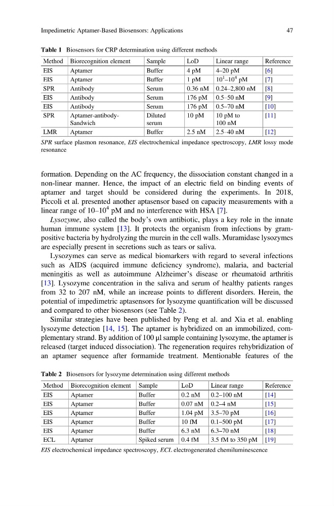
and compared to other biosensors (see Table 2).
Similar strategies have been published by Peng et al. and Xia et al. enabling
lysozyme detection [14, 15]. The aptamer is hybridized on an immobilized, complementary strand. By addition of 100 μl sample containing lysozyme, the aptamer is
released (target induced dissociation). The regeneration requires rehybridization of
an aptamer sequence after formamide treatment. Mentionable features of the
Table 1 Biosensors for CRP determination using different methods
Method Biorecognition element
Sample
LoD
Linear range
Reference
EIS
Aptamer
Buffer
4 pM
4–20 pM
[6]
EIS
Aptamer
Buffer
1 pM
10
1
–10
4 pM
[7]
SPR
Antibody
Serum
0.36 nM 0.24–2,800 nM [8]
EIS
Antibody
Serum
176 pM 0.5–50 nM
[9]
EIS
Antibody
Serum
176 pM 0.5–70 nM
[10]
SPR
Aptamer-antibodySandwich
Diluted
serum
10 pM
10 pM to
100 nM
[11]
LMR
Aptamer
Buffer
2.5 nM
2.5–40 nM
[12]
SPR surface plasmon resonance, EIS electrochemical impedance spectroscopy, LMR lossy mode
resonance
Table 2 Biosensors for lysozyme determination using different methods
Method Biorecognition element Sample
LoD
Linear range
Reference
EIS
Aptamer
Buffer
0.2 nM
0.2–100 nM
[14]
EIS
Aptamer
Buffer
0.07 nM 0.2–4 nM
[15]
EIS
Aptamer
Buffer
1.04 pM 3.5–70 pM
[16]
EIS
Aptamer
Buffer
10 fM
0.1–500 pM
[17]
EIS
Aptamer
Buffer
6.3 nM
6.3–70 nM
[18]
ECL
Aptamer
Spiked serum 0.4 fM
3.5 fM to 350 pM [19]
EIS electrochemical impedance spectroscopy, ECL electrogenerated chemiluminescence
Impedimetric Aptamer-Based Biosensors: Applications
47
non-linear manner. Hence, the impact of an electric field on binding events of
aptamer and target should be considered during the experiments. In 2018,
Piccoli et al. presented another aptasensor based on capacity measurements with a
linear range of 10–10
4 pM and no interference with HSA [7].
Lysozyme, also called the body’s own antibiotic, plays a key role in the innate
human immune system [13]. It protects the organism from infections by grampositive bacteria by hydrolyzing the murein in the cell walls. Muramidase lysozymes
are especially present in secretions such as tears or saliva.
Lysozymes can serve as medical biomarkers with regard to several infections
such as AIDS (acquired immune deficiency syndrome), malaria, and bacterial
meningitis as well as autoimmune Alzheimer’s disease or rheumatoid arthritis
[13]. Lysozyme concentration in the saliva and serum of healthy patients ranges
from 32 to 207 nM, while an increase points to different disorders. Herein, the
potential of impedimetric aptasensors for lysozyme quantification will be discussed
and compared to other biosensors (see Table 2).
Similar strategies have been published by Peng et al. and Xia et al. enabling
lysozyme detection [14, 15]. The aptamer is hybridized on an immobilized, complementary strand. By addition of 100 μl sample containing lysozyme, the aptamer is
released (target induced dissociation). The regeneration requires rehybridization of
an aptamer sequence after formamide treatment. Mentionable features of the
Table 1 Biosensors for CRP determination using different methods
Method Biorecognition element
Sample
LoD
Linear range
Reference
EIS
Aptamer
Buffer
4 pM
4–20 pM
[6]
EIS
Aptamer
Buffer
1 pM
10
1
–10
4 pM
[7]
SPR
Antibody
Serum
0.36 nM 0.24–2,800 nM [8]
EIS
Antibody
Serum
176 pM 0.5–50 nM
[9]
EIS
Antibody
Serum
176 pM 0.5–70 nM
[10]
SPR
Aptamer-antibodySandwich
Diluted
serum
10 pM
10 pM to
100 nM
[11]
LMR
Aptamer
Buffer
2.5 nM
2.5–40 nM
[12]
SPR surface plasmon resonance, EIS electrochemical impedance spectroscopy, LMR lossy mode
resonance
Table 2 Biosensors for lysozyme determination using different methods
Method Biorecognition element Sample
LoD
Linear range
Reference
EIS
Aptamer
Buffer
0.2 nM
0.2–100 nM
[14]
EIS
Aptamer
Buffer
0.07 nM 0.2–4 nM
[15]
EIS
Aptamer
Buffer
1.04 pM 3.5–70 pM
[16]
EIS
Aptamer
Buffer
10 fM
0.1–500 pM
[17]
EIS
Aptamer
Buffer
6.3 nM
6.3–70 nM
[18]
ECL
Aptamer
Spiked serum 0.4 fM
3.5 fM to 350 pM [19]
EIS electrochemical impedance spectroscopy, ECL electrogenerated chemiluminescence
Impedimetric Aptamer-Based Biosensors: Applications
47