

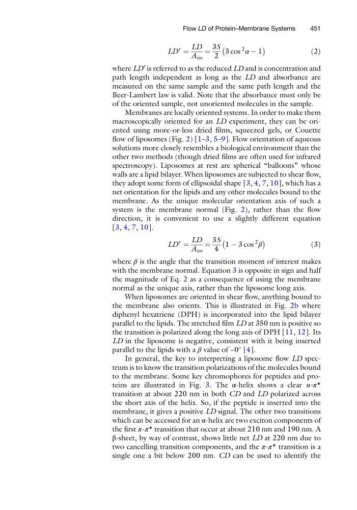
LD
r
¼
LD
A iso
¼
3S
2
3 cos
2
α À 1
À
Á
ð2Þ
where LD
r is referred to as the reduced LD and is concentration and
path length independent as long as the LD and absorbance are
measured on the same sample and the same path length and the
Beer-Lambert law is valid. Note that the absorbance must only be
of the oriented sample, not unoriented molecules in the sample.
Membranes are locally oriented systems. In order to make them
macroscopically oriented for an LD experiment, they can be oriented using more-or-less dried films, squeezed gels, or Couette
flow of liposomes (Fig. 2) [1–3, 5–9]. Flow orientation of aqueous
solutions more closely resembles a biological environment than the
other two methods (though dried films are often used for infrared
spectroscopy). Liposomes at rest are spherical “balloons” whose
walls are a lipid bilayer. When liposomes are subjected to shear flow,
they adopt some form of ellipsoidal shape [3, 4, 7, 10], which has a
net orientation for the lipids and any other molecules bound to the
membrane. As the unique molecular orientation axis of such a
system is the membrane normal (Fig. 2), rather than the flow
direction, it is convenient to use a slightly different equation
[3, 4, 7, 10].
LD
r
¼
LD
A iso
¼
3S
4
1 À 3 cos
2
β
À
Á
ð3Þ
where β is the angle that the transition moment of interest makes
with the membrane normal. Equation 3 is opposite in sign and half
the magnitude of Eq. 2 as a consequence of using the membrane
normal as the unique axis, rather than the liposome long axis.
When liposomes are oriented in shear flow, anything bound to
the membrane also orients. This is illustrated in Fig. 2b where
diphenyl hexatriene (DPH) is incorporated into the lipid bilayer
parallel to the lipids. The stretched film LD at 350 nm is positive so
the transition is polarized along the long axis of DPH [11, 12]. Its
LD in the liposome is negative, consistent with it being inserted
parallel to the lipids with a β value of ~0
[4].
In general, the key to interpreting a liposome flow LD spectrum is to know the transition polarizations of the molecules bound
to the membrane. Some key chromophores for peptides and proteins are illustrated in Fig. 3. The α-helix shows a clear n-π*
transition at about 220 nm in both CD and LD polarized across
the short axis of the helix. So, if the peptide is inserted into the
membrane, it gives a positive LD signal. The other two transitions
which can be accessed for an α-helix are two exciton components of
the first π-π* transition that occur at about 210 nm and 190 nm. A
β-sheet, by way of contrast, shows little net LD at 220 nm due to
two cancelling transition components, and the π-π* transition is a
single one a bit below 200 nm. CD can be used to identify the
Flow LD of Protein–Membrane Systems
451
r
¼
LD
A iso
¼
3S
2
3 cos
2
α À 1
À
Á
ð2Þ
where LD
r is referred to as the reduced LD and is concentration and
path length independent as long as the LD and absorbance are
measured on the same sample and the same path length and the
Beer-Lambert law is valid. Note that the absorbance must only be
of the oriented sample, not unoriented molecules in the sample.
Membranes are locally oriented systems. In order to make them
macroscopically oriented for an LD experiment, they can be oriented using more-or-less dried films, squeezed gels, or Couette
flow of liposomes (Fig. 2) [1–3, 5–9]. Flow orientation of aqueous
solutions more closely resembles a biological environment than the
other two methods (though dried films are often used for infrared
spectroscopy). Liposomes at rest are spherical “balloons” whose
walls are a lipid bilayer. When liposomes are subjected to shear flow,
they adopt some form of ellipsoidal shape [3, 4, 7, 10], which has a
net orientation for the lipids and any other molecules bound to the
membrane. As the unique molecular orientation axis of such a
system is the membrane normal (Fig. 2), rather than the flow
direction, it is convenient to use a slightly different equation
[3, 4, 7, 10].
LD
r
¼
LD
A iso
¼
3S
4
1 À 3 cos
2
β
À
Á
ð3Þ
where β is the angle that the transition moment of interest makes
with the membrane normal. Equation 3 is opposite in sign and half
the magnitude of Eq. 2 as a consequence of using the membrane
normal as the unique axis, rather than the liposome long axis.
When liposomes are oriented in shear flow, anything bound to
the membrane also orients. This is illustrated in Fig. 2b where
diphenyl hexatriene (DPH) is incorporated into the lipid bilayer
parallel to the lipids. The stretched film LD at 350 nm is positive so
the transition is polarized along the long axis of DPH [11, 12]. Its
LD in the liposome is negative, consistent with it being inserted
parallel to the lipids with a β value of ~0
[4].
In general, the key to interpreting a liposome flow LD spectrum is to know the transition polarizations of the molecules bound
to the membrane. Some key chromophores for peptides and proteins are illustrated in Fig. 3. The α-helix shows a clear n-π*
transition at about 220 nm in both CD and LD polarized across
the short axis of the helix. So, if the peptide is inserted into the
membrane, it gives a positive LD signal. The other two transitions
which can be accessed for an α-helix are two exciton components of
the first π-π* transition that occur at about 210 nm and 190 nm. A
β-sheet, by way of contrast, shows little net LD at 220 nm due to
two cancelling transition components, and the π-π* transition is a
single one a bit below 200 nm. CD can be used to identify the
Flow LD of Protein–Membrane Systems
451