

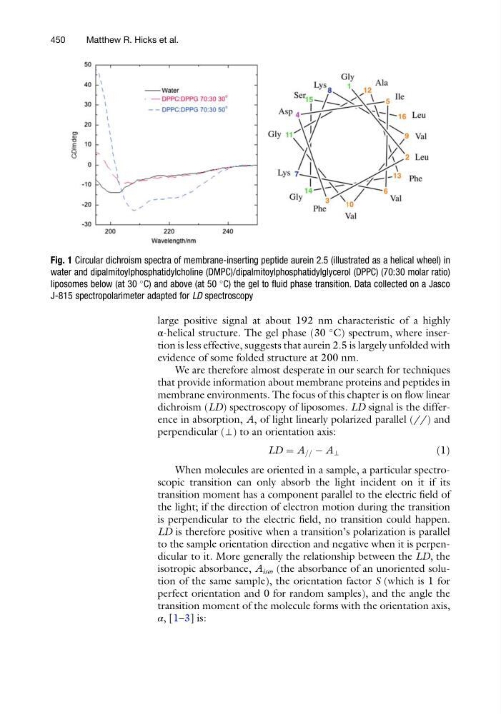
large positive signal at about 192 nm characteristic of a highly
α-helical structure. The gel phase (30
C) spectrum, where insertion is less effective, suggests that aurein 2.5 is largely unfolded with
evidence of some folded structure at 200 nm.
We are therefore almost desperate in our search for techniques
that provide information about membrane proteins and peptides in
membrane environments. The focus of this chapter is on flow linear
dichroism (LD) spectroscopy of liposomes. LD signal is the difference in absorption, A, of light linearly polarized parallel (//) and
perpendicular (⊥) to an orientation axis:
LD ¼ A == À A ⊥
ð1Þ
When molecules are oriented in a sample, a particular spectroscopic transition can only absorb the light incident on it if its
transition moment has a component parallel to the electric field of
the light; if the direction of electron motion during the transition
is perpendicular to the electric field, no transition could happen.
LD is therefore positive when a transition’s polarization is parallel
to the sample orientation direction and negative when it is perpendicular to it. More generally the relationship between the LD, the
isotropic absorbance, A iso , (the absorbance of an unoriented solution of the same sample), the orientation factor S (which is 1 for
perfect orientation and 0 for random samples), and the angle the
transition moment of the molecule forms with the orientation axis,
α, [1–3] is:
Fig. 1 Circular dichroism spectra of membrane-inserting peptide aurein 2.5 (illustrated as a helical wheel) in
water and dipalmitoylphosphatidylcholine (DMPC)/dipalmitoylphosphatidylglycerol (DPPC) (70:30 molar ratio)
liposomes below (at 30
C) and above (at 50
C) the gel to fluid phase transition. Data collected on a Jasco
J-815 spectropolarimeter adapted for LD spectroscopy
450
Matthew R. Hicks et al.
α-helical structure. The gel phase (30
C) spectrum, where insertion is less effective, suggests that aurein 2.5 is largely unfolded with
evidence of some folded structure at 200 nm.
We are therefore almost desperate in our search for techniques
that provide information about membrane proteins and peptides in
membrane environments. The focus of this chapter is on flow linear
dichroism (LD) spectroscopy of liposomes. LD signal is the difference in absorption, A, of light linearly polarized parallel (//) and
perpendicular (⊥) to an orientation axis:
LD ¼ A == À A ⊥
ð1Þ
When molecules are oriented in a sample, a particular spectroscopic transition can only absorb the light incident on it if its
transition moment has a component parallel to the electric field of
the light; if the direction of electron motion during the transition
is perpendicular to the electric field, no transition could happen.
LD is therefore positive when a transition’s polarization is parallel
to the sample orientation direction and negative when it is perpendicular to it. More generally the relationship between the LD, the
isotropic absorbance, A iso , (the absorbance of an unoriented solution of the same sample), the orientation factor S (which is 1 for
perfect orientation and 0 for random samples), and the angle the
transition moment of the molecule forms with the orientation axis,
α, [1–3] is:
Fig. 1 Circular dichroism spectra of membrane-inserting peptide aurein 2.5 (illustrated as a helical wheel) in
water and dipalmitoylphosphatidylcholine (DMPC)/dipalmitoylphosphatidylglycerol (DPPC) (70:30 molar ratio)
liposomes below (at 30
C) and above (at 50
C) the gel to fluid phase transition. Data collected on a Jasco
J-815 spectropolarimeter adapted for LD spectroscopy
450
Matthew R. Hicks et al.