

secondary structure of a peptide. So, if a peptide binds to a liposome, it will have LD signals for its transitions whose signs and
magnitudes depend on the degree of orientation, S, and the orientation on the membrane (Eq. 3). S is typically of the order of 0.03.
If a helix is inserted into the membrane, then its 208 nm
transition will be negative and the 195 nm and 222 nm transitions
positive. However, the 208 nm transition is overlaid by the others
and never actually becomes negative. Figure 4 summarizes the
possibilities for α-helices and β-strands. Thus, the sign of peptide
LD indicates its orientation with respect to the membrane normal.
Other technique will need to be used to determine whether a
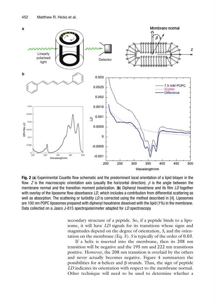
Fig. 2 (a) Experimental Couette flow schematic and the predominant local orientation of a lipid bilayer in the
flow. Z is the macroscopic orientation axis (usually the horizontal direction). β is the angle between the
membrane normal and the transition moment polarization. (b) Diphenyl hexatriene and its film LD together
with overlay of the liposome flow absorbance LD, which includes a contribution from differential scattering as
well as absorption. The scattering or turbidity LD is corrected using the method described in [4]. Liposomes
are 100 nm POPC liposomes prepared with diphenyl hexatriene dissolved with the lipid (1%) in the membrane.
Data collected on a Jasco J-815 spectropolarimeter adapted for LD spectroscopy
452
Matthew R. Hicks et al.
magnitudes depend on the degree of orientation, S, and the orientation on the membrane (Eq. 3). S is typically of the order of 0.03.
If a helix is inserted into the membrane, then its 208 nm
transition will be negative and the 195 nm and 222 nm transitions
positive. However, the 208 nm transition is overlaid by the others
and never actually becomes negative. Figure 4 summarizes the
possibilities for α-helices and β-strands. Thus, the sign of peptide
LD indicates its orientation with respect to the membrane normal.
Other technique will need to be used to determine whether a
Fig. 2 (a) Experimental Couette flow schematic and the predominant local orientation of a lipid bilayer in the
flow. Z is the macroscopic orientation axis (usually the horizontal direction). β is the angle between the
membrane normal and the transition moment polarization. (b) Diphenyl hexatriene and its film LD together
with overlay of the liposome flow absorbance LD, which includes a contribution from differential scattering as
well as absorption. The scattering or turbidity LD is corrected using the method described in [4]. Liposomes
are 100 nm POPC liposomes prepared with diphenyl hexatriene dissolved with the lipid (1%) in the membrane.
Data collected on a Jasco J-815 spectropolarimeter adapted for LD spectroscopy
452
Matthew R. Hicks et al.