

Mechanism in Protein Science by Alan Fersht [8], or other similar
enzyme kinetics texts.
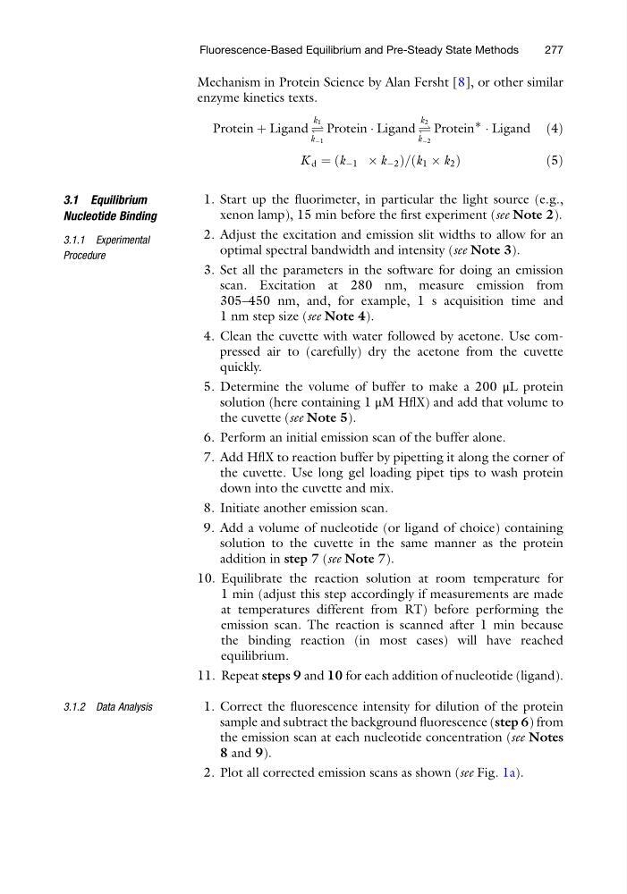
Protein þ Ligand Ð
k 1
k À1
Protein Á Ligand Ð
k 2
k À2
Protein
∗
Á Ligand ð4Þ
K d ¼ k À1 Â k À2
ð
Þ = k 1 Â k 2
ð
Þ
ð5Þ
3.1 Equilibrium
Nucleotide Binding
3.1.1 Experimental
Procedure
1. Start up the fluorimeter, in particular the light source (e.g.,
xenon lamp), 15 min before the first experiment (see Note 2).
2. Adjust the excitation and emission slit widths to allow for an
optimal spectral bandwidth and intensity (see Note 3).
3. Set all the parameters in the software for doing an emission
scan. Excitation at 280 nm, measure emission from
305–450 nm, and, for example, 1 s acquisition time and
1 nm step size (see Note 4).
4. Clean the cuvette with water followed by acetone. Use compressed air to (carefully) dry the acetone from the cuvette
quickly.
5. Determine the volume of buffer to make a 200 μL protein
solution (here containing 1 μM HflX) and add that volume to
the cuvette (see Note 5).
6. Perform an initial emission scan of the buffer alone.
7. Add HflX to reaction buffer by pipetting it along the corner of
the cuvette. Use long gel loading pipet tips to wash protein
down into the cuvette and mix.
8. Initiate another emission scan.
9. Add a volume of nucleotide (or ligand of choice) containing
solution to the cuvette in the same manner as the protein
addition in step 7 (see Note 7).
10. Equilibrate the reaction solution at room temperature for
1 min (adjust this step accordingly if measurements are made
at temperatures different from RT) before performing the
emission scan. The reaction is scanned after 1 min because
the binding reaction (in most cases) will have reached
equilibrium.
11. Repeat steps 9 and 10 for each addition of nucleotide (ligand).
3.1.2 Data Analysis
1. Correct the fluorescence intensity for dilution of the protein
sample and subtract the background fluorescence (step 6) from
the emission scan at each nucleotide concentration (see Notes
8 and 9).
2. Plot all corrected emission scans as shown (see Fig. 1a).
Fluorescence-Based Equilibrium and Pre-Steady State Methods
277
enzyme kinetics texts.
Protein þ Ligand Ð
k 1
k À1
Protein Á Ligand Ð
k 2
k À2
Protein
∗
Á Ligand ð4Þ
K d ¼ k À1 Â k À2
ð
Þ = k 1 Â k 2
ð
Þ
ð5Þ
3.1 Equilibrium
Nucleotide Binding
3.1.1 Experimental
Procedure
1. Start up the fluorimeter, in particular the light source (e.g.,
xenon lamp), 15 min before the first experiment (see Note 2).
2. Adjust the excitation and emission slit widths to allow for an
optimal spectral bandwidth and intensity (see Note 3).
3. Set all the parameters in the software for doing an emission
scan. Excitation at 280 nm, measure emission from
305–450 nm, and, for example, 1 s acquisition time and
1 nm step size (see Note 4).
4. Clean the cuvette with water followed by acetone. Use compressed air to (carefully) dry the acetone from the cuvette
quickly.
5. Determine the volume of buffer to make a 200 μL protein
solution (here containing 1 μM HflX) and add that volume to
the cuvette (see Note 5).
6. Perform an initial emission scan of the buffer alone.
7. Add HflX to reaction buffer by pipetting it along the corner of
the cuvette. Use long gel loading pipet tips to wash protein
down into the cuvette and mix.
8. Initiate another emission scan.
9. Add a volume of nucleotide (or ligand of choice) containing
solution to the cuvette in the same manner as the protein
addition in step 7 (see Note 7).
10. Equilibrate the reaction solution at room temperature for
1 min (adjust this step accordingly if measurements are made
at temperatures different from RT) before performing the
emission scan. The reaction is scanned after 1 min because
the binding reaction (in most cases) will have reached
equilibrium.
11. Repeat steps 9 and 10 for each addition of nucleotide (ligand).
3.1.2 Data Analysis
1. Correct the fluorescence intensity for dilution of the protein
sample and subtract the background fluorescence (step 6) from
the emission scan at each nucleotide concentration (see Notes
8 and 9).
2. Plot all corrected emission scans as shown (see Fig. 1a).
Fluorescence-Based Equilibrium and Pre-Steady State Methods
277