

formed, the surface can be used to monitor more complex interactions. Figure 3 shows how we used these layers to study the interaction of a heparin-binding protein with end-biotinylated dp-24
heparin. Following bilayer formation with 2 mol% Biotinyl-Cap PE
lipids, streptavidin was added to the layer followed by heparin. The
addition of heparin gives a characteristically large change in dissipation caused by the viscoelastic nature of the highly solvated heparin
chains arranged perpendicular to the surface as compared to the
small dissipation change caused by streptavidin, which forms a
compact layer. Subsequent interaction of a heparin-binding protein
causes a collapse of the heparin layer caused by heparin crosslinking. Interaction of the protein to the heparin is seen by the
change in frequency, which is the result of mass addition, but the
sudden decrease in dissipation means that there is a significant
change in surface viscoelasticity caused by a collapse of the heparin
chains. This phenomenon can only be interpreted using dissipation
measurements from QCM-D as the collapse of the heparin layer is
hidden when using other purely mass sensing techniques, such as
surface plasmon resonance (SPR), and this highlights the unique
advantages of this methodology.
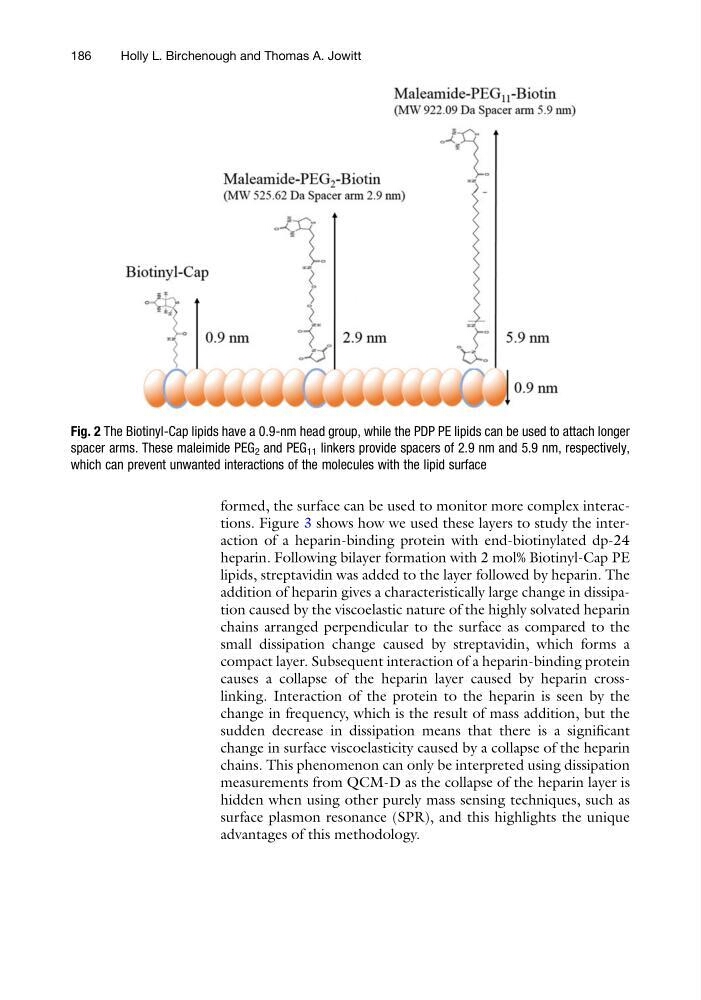
Fig. 2 The Biotinyl-Cap lipids have a 0.9-nm head group, while the PDP PE lipids can be used to attach longer
spacer arms. These maleimide PEG 2 and PEG 11 linkers provide spacers of 2.9 nm and 5.9 nm, respectively,
which can prevent unwanted interactions of the molecules with the lipid surface
186
Holly L. Birchenough and Thomas A. Jowitt
heparin. Following bilayer formation with 2 mol% Biotinyl-Cap PE
lipids, streptavidin was added to the layer followed by heparin. The
addition of heparin gives a characteristically large change in dissipation caused by the viscoelastic nature of the highly solvated heparin
chains arranged perpendicular to the surface as compared to the
small dissipation change caused by streptavidin, which forms a
compact layer. Subsequent interaction of a heparin-binding protein
causes a collapse of the heparin layer caused by heparin crosslinking. Interaction of the protein to the heparin is seen by the
change in frequency, which is the result of mass addition, but the
sudden decrease in dissipation means that there is a significant
change in surface viscoelasticity caused by a collapse of the heparin
chains. This phenomenon can only be interpreted using dissipation
measurements from QCM-D as the collapse of the heparin layer is
hidden when using other purely mass sensing techniques, such as
surface plasmon resonance (SPR), and this highlights the unique
advantages of this methodology.
Fig. 2 The Biotinyl-Cap lipids have a 0.9-nm head group, while the PDP PE lipids can be used to attach longer
spacer arms. These maleimide PEG 2 and PEG 11 linkers provide spacers of 2.9 nm and 5.9 nm, respectively,
which can prevent unwanted interactions of the molecules with the lipid surface
186
Holly L. Birchenough and Thomas A. Jowitt