

Here we describe the method for the production of functionalized lipid bilayers on QCM-D SiO 2 sensor surfaces, which have the
capability of further immobilizing molecules of interest for analysis.
We describe two methods of functionalization: first, using a biotinylated lipid for direct immobilization of biotin-binding avidin/
streptavidin to the lipid layer; and second, using a lipid containing a
disulfide bond within a head group that can be reduced to expose a
free sulfhydryl for the immobilization of avidin/streptavidin via
maleimide–biotin linkers (Fig. 2). This extended approach allows
for the addition of spacer arms to increase the flexibility of the
immobilized protein. Once functionalized bilayers have been
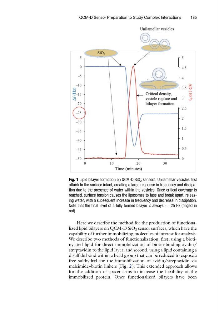
Fig. 1 Lipid bilayer formation on QCM-D SiO 2 sensors. Unilamellar vesicles first
attach to the surface intact, creating a large response in frequency and dissipation due to the presence of water within the vesicles. Once critical coverage is
reached, surface tension causes the liposomes to fuse and break apart, releasing water, with a subsequent increase in frequency and decrease in dissipation.
Note that the final level of a fully formed bilayer is always ~À25 Hz (ringed in
red)
QCM-D Sensor Preparation to Study Complex Interactions
185
capability of further immobilizing molecules of interest for analysis.
We describe two methods of functionalization: first, using a biotinylated lipid for direct immobilization of biotin-binding avidin/
streptavidin to the lipid layer; and second, using a lipid containing a
disulfide bond within a head group that can be reduced to expose a
free sulfhydryl for the immobilization of avidin/streptavidin via
maleimide–biotin linkers (Fig. 2). This extended approach allows
for the addition of spacer arms to increase the flexibility of the
immobilized protein. Once functionalized bilayers have been
Fig. 1 Lipid bilayer formation on QCM-D SiO 2 sensors. Unilamellar vesicles first
attach to the surface intact, creating a large response in frequency and dissipation due to the presence of water within the vesicles. Once critical coverage is
reached, surface tension causes the liposomes to fuse and break apart, releasing water, with a subsequent increase in frequency and decrease in dissipation.
Note that the final level of a fully formed bilayer is always ~À25 Hz (ringed in
red)
QCM-D Sensor Preparation to Study Complex Interactions
185