

classified it from the “recretohalophytes” plants (Dassanayake and Larkin 2017).
However, leaf primordia of branchlet apices of this halophyte have been found to
bear external salt glands at different developmental stages. Further light and electron
microscopic observations revealed that all these stages from initiation of salt glands
up to their final differentiation also occur in fully-expanded mature leaves. In
T. atriculata (aphylla), unlike the species of other families, external leaf glands are
not permanent on leaves, but by the time some glands mature and disintegrate, some
others initiate and develop (Bosabalidis 1992). It was recognized that salt glands
vary greatly among plants and can be divided into multicellular salt glands, bicellular
salt glands, and salt bladders according to their structure (Ma et al. 2011). For
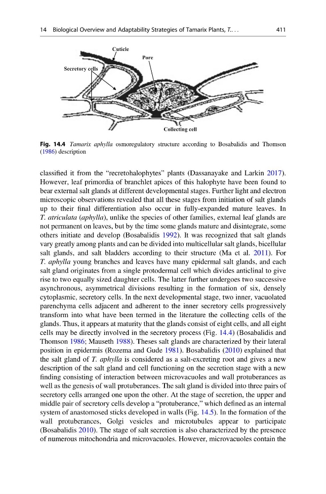
T. aphylla young branches and leaves have many epidermal salt glands, and each
salt gland originates from a single protodermal cell which divides anticlinal to give
rise to two equally sized daughter cells. The latter further undergoes two successive
asynchronous, asymmetrical divisions resulting in the formation of six, densely
cytoplasmic, secretory cells. In the next developmental stage, two inner, vacuolated
parenchyma cells adjacent and adherent to the inner secretory cells progressively
transform into what have been termed in the literature the collecting cells of the
glands. Thus, it appears at maturity that the glands consist of eight cells, and all eight
cells may be directly involved in the secretory process (Fig. 14.4) (Bosabalidis and
Thomson 1986; Mauseth 1988). Theses salt glands are characterized by their lateral
position in epidermis (Rozema and Gude 1981). Bosabalidis (2010) explained that
the salt gland of T. aphylla is considered as a salt-excreting root and gives a new
description of the salt gland and cell functioning on the secretion stage with a new
finding consisting of interaction between microvacuoles and wall protuberances as
well as the genesis of wall protuberances. The salt gland is divided into three pairs of
secretory cells arranged one upon the other. At the stage of secretion, the upper and
middle pair of secretory cells develop a “protuberance,” which defined as an internal
system of anastomosed sticks developed in walls (Fig. 14.5). In the formation of the
wall protuberances, Golgi vesicles and microtubules appear to participate
(Bosabalidis 2010). The stage of salt secretion is also characterized by the presence
of numerous mitochondria and microvacuoles. However, microvacuoles contain the
Collecting cell
Secretory cells
Cuticle
Pore
Fig. 14.4 Tamarix aphylla osmoregulatory structure according to Bosabalidis and Thomson
(1986) description
14 Biological Overview and Adaptability Strategies of Tamarix Plants, T.. . .
411
However, leaf primordia of branchlet apices of this halophyte have been found to
bear external salt glands at different developmental stages. Further light and electron
microscopic observations revealed that all these stages from initiation of salt glands
up to their final differentiation also occur in fully-expanded mature leaves. In
T. atriculata (aphylla), unlike the species of other families, external leaf glands are
not permanent on leaves, but by the time some glands mature and disintegrate, some
others initiate and develop (Bosabalidis 1992). It was recognized that salt glands
vary greatly among plants and can be divided into multicellular salt glands, bicellular
salt glands, and salt bladders according to their structure (Ma et al. 2011). For
T. aphylla young branches and leaves have many epidermal salt glands, and each
salt gland originates from a single protodermal cell which divides anticlinal to give
rise to two equally sized daughter cells. The latter further undergoes two successive
asynchronous, asymmetrical divisions resulting in the formation of six, densely
cytoplasmic, secretory cells. In the next developmental stage, two inner, vacuolated
parenchyma cells adjacent and adherent to the inner secretory cells progressively
transform into what have been termed in the literature the collecting cells of the
glands. Thus, it appears at maturity that the glands consist of eight cells, and all eight
cells may be directly involved in the secretory process (Fig. 14.4) (Bosabalidis and
Thomson 1986; Mauseth 1988). Theses salt glands are characterized by their lateral
position in epidermis (Rozema and Gude 1981). Bosabalidis (2010) explained that
the salt gland of T. aphylla is considered as a salt-excreting root and gives a new
description of the salt gland and cell functioning on the secretion stage with a new
finding consisting of interaction between microvacuoles and wall protuberances as
well as the genesis of wall protuberances. The salt gland is divided into three pairs of
secretory cells arranged one upon the other. At the stage of secretion, the upper and
middle pair of secretory cells develop a “protuberance,” which defined as an internal
system of anastomosed sticks developed in walls (Fig. 14.5). In the formation of the
wall protuberances, Golgi vesicles and microtubules appear to participate
(Bosabalidis 2010). The stage of salt secretion is also characterized by the presence
of numerous mitochondria and microvacuoles. However, microvacuoles contain the
Collecting cell
Secretory cells
Cuticle
Pore
Fig. 14.4 Tamarix aphylla osmoregulatory structure according to Bosabalidis and Thomson
(1986) description
14 Biological Overview and Adaptability Strategies of Tamarix Plants, T.. . .
411