

glycosylated forms to enhance their water solubility increasing their bioavailability. It also makes the
flavonoids less reactive and allows prevention of cytoplasmic damage (Cuyckens & Claeys, 2004; Slámová,
Kapešová & Kapešová 2018;.
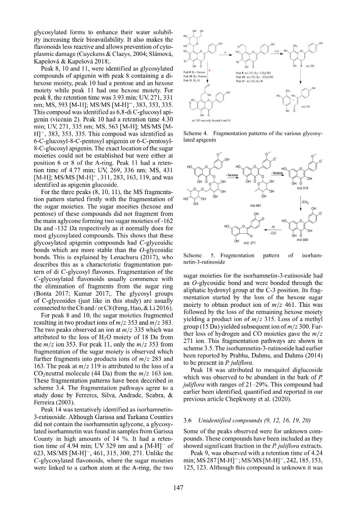
Peak 8, 10 and 11, were identified as glycosylated
compounds of apigenin with peak 8 containing a dihexose moiety, peak 10 had a pentose and an hexose
moiety while peak 11 had one hexose moiety. For
peak 8, the retention time was 3.93 min; UV, 271, 331
nm; MS, 593 [M-H]; MS/MS [M-H]
− , 383, 353, 335.
This compoud was identified as 6,8-di C-glucosyl apigenin (vicenin 2). Peak 10 had a retention time 4.30
min; UV, 271, 335 nm; MS, 563 [M-H]; MS/MS [MH]
− , 383, 353, 335. This compoud was identified as
6-C-glucosyl-8-C-pentosyl apigenin or 6-C-pentosyl8-C-glucosyl apigenin. The exact location of the sugar
moieties could not be established but were either at
position 6 or 8 of the A-ring. Peak 11 had a retention time of 4.77 min; UV, 269, 336 nm; MS, 431
[M-H]; MS/MS [M-H]
− , 311, 283, 163, 119, and was
identified as apigenin glucoside.
For the three peaks (8, 10, 11), the MS fragmentation pattern started firstly with the fragmentation of
the sugar moieties. The sugar moeities (hexose and
pentose) of these compounds did not fragment from
the main aglycone forming two sugar moieties of -162
Da and -132 Da respectively as it normally does for
most glycosylated compounds. This shows that these
glycosylated apigenin compounds had C-glycosidic
bonds which are more stable than the O-glycosidic
bonds. This is explained by Lenachuru (2017), who
describes this as a characteristic fragmentation pattern of di C-glycosyl flavones. Fragmentation of the
C-glycosylated flavonoids usually commence with
the elimination of fragments from the sugar ring
(Bonta 2017; Kumar 2017;. The glycosyl groups
of C-glycosides (just like in this study) are usually
connected to the C6 and / or C8 (Feng, Hao, & Li 2016).
For peak 8 and 10, the sugar moieties fragmented
resulting in two product ions of m/z 353 and m/z 383.
The two peaks observed an ion at m/z 335 which was
attributed to the loss of H 2 O moiety of 18 Da from
the m/z ion 353. For peak 11, only the m/z 353 from
fragmentation of the sugar moiety is observed which
further fragments into products ions of m/z 283 and
163. The peak at m/z 119 is attributed to the loss of a
CO 2 neutral molecule (44 Da) from the m/z 163 ion.
These fragmentation patterns have been deccribed in
scheme 3.4. The fragmentation pathways agree to a
study done by Ferreres, Silva, Andrade, Seabra, &
Ferreira (2003).
Peak 14 was tentatively identified as isorhamnetin3-rutinoside. Although Garissa and Turkana Counties
did not contain the isorhamnetin aglycone, a glycosylated isorhamnetin was found in samples from Garissa
County in high amounts of 14 %. It had a retention time of 4.94 min; UV 329 nm and a [M-H]
− of
623, MS/MS [M-H]
− , 461, 315, 300, 271. Unlike the
C-glycosylated flavonoids, where the sugar moieties
were linked to a carbon atom at the A-ring, the two
Scheme 4. Fragmentation patterns of the various glycosylated apigenin
Scheme
5. Fragmentation
pattern
of
isorhamnetin-3-rutinoside
sugar moieties for the isorhamnetin-3-rutinoside had
an O-glycosidic bond and were bonded through the
aliphatic hydroxyl group at the C-3 position. Its fragmentation started by the loss of the hexose sugar
moiety to obtain product ion of m/z 461. This was
followed by the loss of the remaining hexose moiety
yielding a product ion of m/z 315. Loss of a methyl
group (15 Da) yielded subsequent ion of m/z 300. Further loss of hydrogen and CO moieties gave the m/z
271 ion. This fragmentation pathways are shown in
scheme 3.5. The isorhamnetin-3-rutinoside had earlier
been reported by Prabha, Dahms, and Dahms (2014)
to be present in P. juliflora.
Peak 18 was attributed to mesquitol diglucoside
which was observed to be abundant in the bark of P.
juliflora with ranges of 21–29%. This compound had
earlier been identified, quantified and reported in our
previous article Chepkwony et al. (2020).
3.6 Unidentified compounds (9, 12, 16, 19, 20)
Some of the peaks observed were for unknown compounds. These compounds have been included as they
showed significant fraction in the P. juliflora extracts.
Peak 9, was observed with a retention time of 4.24
min; MS 287 [M-H]
− ; MS/MS [M-H]
− , 242, 185, 153,
125, 123. Although this compound is unknown it was
147
flavonoids less reactive and allows prevention of cytoplasmic damage (Cuyckens & Claeys, 2004; Slámová,
Kapešová & Kapešová 2018;.
Peak 8, 10 and 11, were identified as glycosylated
compounds of apigenin with peak 8 containing a dihexose moiety, peak 10 had a pentose and an hexose
moiety while peak 11 had one hexose moiety. For
peak 8, the retention time was 3.93 min; UV, 271, 331
nm; MS, 593 [M-H]; MS/MS [M-H]
− , 383, 353, 335.
This compoud was identified as 6,8-di C-glucosyl apigenin (vicenin 2). Peak 10 had a retention time 4.30
min; UV, 271, 335 nm; MS, 563 [M-H]; MS/MS [MH]
− , 383, 353, 335. This compoud was identified as
6-C-glucosyl-8-C-pentosyl apigenin or 6-C-pentosyl8-C-glucosyl apigenin. The exact location of the sugar
moieties could not be established but were either at
position 6 or 8 of the A-ring. Peak 11 had a retention time of 4.77 min; UV, 269, 336 nm; MS, 431
[M-H]; MS/MS [M-H]
− , 311, 283, 163, 119, and was
identified as apigenin glucoside.
For the three peaks (8, 10, 11), the MS fragmentation pattern started firstly with the fragmentation of
the sugar moieties. The sugar moeities (hexose and
pentose) of these compounds did not fragment from
the main aglycone forming two sugar moieties of -162
Da and -132 Da respectively as it normally does for
most glycosylated compounds. This shows that these
glycosylated apigenin compounds had C-glycosidic
bonds which are more stable than the O-glycosidic
bonds. This is explained by Lenachuru (2017), who
describes this as a characteristic fragmentation pattern of di C-glycosyl flavones. Fragmentation of the
C-glycosylated flavonoids usually commence with
the elimination of fragments from the sugar ring
(Bonta 2017; Kumar 2017;. The glycosyl groups
of C-glycosides (just like in this study) are usually
connected to the C6 and / or C8 (Feng, Hao, & Li 2016).
For peak 8 and 10, the sugar moieties fragmented
resulting in two product ions of m/z 353 and m/z 383.
The two peaks observed an ion at m/z 335 which was
attributed to the loss of H 2 O moiety of 18 Da from
the m/z ion 353. For peak 11, only the m/z 353 from
fragmentation of the sugar moiety is observed which
further fragments into products ions of m/z 283 and
163. The peak at m/z 119 is attributed to the loss of a
CO 2 neutral molecule (44 Da) from the m/z 163 ion.
These fragmentation patterns have been deccribed in
scheme 3.4. The fragmentation pathways agree to a
study done by Ferreres, Silva, Andrade, Seabra, &
Ferreira (2003).
Peak 14 was tentatively identified as isorhamnetin3-rutinoside. Although Garissa and Turkana Counties
did not contain the isorhamnetin aglycone, a glycosylated isorhamnetin was found in samples from Garissa
County in high amounts of 14 %. It had a retention time of 4.94 min; UV 329 nm and a [M-H]
− of
623, MS/MS [M-H]
− , 461, 315, 300, 271. Unlike the
C-glycosylated flavonoids, where the sugar moieties
were linked to a carbon atom at the A-ring, the two
Scheme 4. Fragmentation patterns of the various glycosylated apigenin
Scheme
5. Fragmentation
pattern
of
isorhamnetin-3-rutinoside
sugar moieties for the isorhamnetin-3-rutinoside had
an O-glycosidic bond and were bonded through the
aliphatic hydroxyl group at the C-3 position. Its fragmentation started by the loss of the hexose sugar
moiety to obtain product ion of m/z 461. This was
followed by the loss of the remaining hexose moiety
yielding a product ion of m/z 315. Loss of a methyl
group (15 Da) yielded subsequent ion of m/z 300. Further loss of hydrogen and CO moieties gave the m/z
271 ion. This fragmentation pathways are shown in
scheme 3.5. The isorhamnetin-3-rutinoside had earlier
been reported by Prabha, Dahms, and Dahms (2014)
to be present in P. juliflora.
Peak 18 was attributed to mesquitol diglucoside
which was observed to be abundant in the bark of P.
juliflora with ranges of 21–29%. This compound had
earlier been identified, quantified and reported in our
previous article Chepkwony et al. (2020).
3.6 Unidentified compounds (9, 12, 16, 19, 20)
Some of the peaks observed were for unknown compounds. These compounds have been included as they
showed significant fraction in the P. juliflora extracts.
Peak 9, was observed with a retention time of 4.24
min; MS 287 [M-H]
− ; MS/MS [M-H]
− , 242, 185, 153,
125, 123. Although this compound is unknown it was
147