

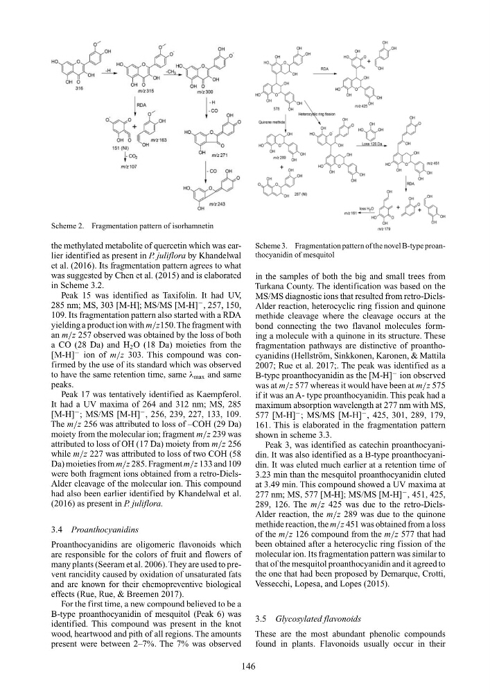
Scheme 2. Fragmentation pattern of isorhamnetin
the methylated metabolite of quercetin which was earlier identified as present in P. juliflora by Khandelwal
et al. (2016). Its fragmentation pattern agrees to what
was suggested by Chen et al. (2015) and is elaborated
in Scheme 3.2.
Peak 15 was identified as Taxifolin. It had UV,
285 nm; MS, 303 [M-H]; MS/MS [M-H]
− , 257, 150,
109. Its fragmentation pattern also started with a RDA
yielding a product ion with m/z150. The fragment with
an m/z 257 observed was obtained by the loss of both
a CO (28 Da) and H 2 O (18 Da) moieties from the
[M-H]
− ion of m/z 303. This compound was confirmed by the use of its standard which was observed
to have the same retention time, same λ max and same
peaks.
Peak 17 was tentatively identified as Kaempferol.
It had a UV maxima of 264 and 312 nm; MS, 285
[M-H]
− ; MS/MS [M-H]
− , 256, 239, 227, 133, 109.
The m/z 256 was attributed to loss of –COH (29 Da)
moiety from the molecular ion; fragment m/z 239 was
attributed to loss of OH (17 Da) moiety from m/z 256
while m/z 227 was attributed to loss of two COH (58
Da) moieties from m/z 285. Fragment m/z 133 and 109
were both fragment ions obtained from a retro-DielsAlder cleavage of the molecular ion. This compound
had also been earlier identified by Khandelwal et al.
(2016) as present in P. juliflora.
3.4 Proanthocyanidins
Proanthocyanidins are oligomeric flavonoids which
are responsible for the colors of fruit and flowers of
many plants (Seeram et al. 2006). They are used to prevent rancidity caused by oxidation of unsaturated fats
and are known for their chemopreventive biological
effects (Rue, Rue, & Breemen 2017).
For the first time, a new compound believed to be a
B-type proanthocyanidin of mesquitol (Peak 6) was
identified. This compound was present in the knot
wood, heartwood and pith of all regions. The amounts
present were between 2–7%. The 7% was observed
Scheme 3. Fragmentation pattern of the novel B-type proanthocyanidin of mesquitol
in the samples of both the big and small trees from
Turkana County. The identification was based on the
MS/MS diagnostic ions that resulted from retro-DielsAlder reaction, heterocyclic ring fission and quinone
methide cleavage where the cleavage occurs at the
bond connecting the two flavanol molecules forming a molecule with a quinone in its structure. These
fragmentation pathways are distinctive of proanthocyanidins (Hellström, Sinkkonen, Karonen, & Mattila
2007; Rue et al. 2017;. The peak was identified as a
B-type proanthocyanidin as the [M-H]
− ion observed
was at m/z 577 whereas it would have been at m/z 575
if it was an A- type proanthocyanidin. This peak had a
maximum absorption wavelength at 277 nm with MS,
577 [M-H]
− ; MS/MS [M-H]
− , 425, 301, 289, 179,
161. This is elaborated in the fragmentation pattern
shown in scheme 3.3.
Peak 3, was identified as catechin proanthocyanidin. It was also identified as a B-type proanthocyanidin. It was eluted much earlier at a retention time of
3.23 min than the mesquitol proanthocyanidin eluted
at 3.49 min. This compound showed a UV maxima at
277 nm; MS, 577 [M-H]; MS/MS [M-H]
− , 451, 425,
289, 126. The m/z 425 was due to the retro-DielsAlder reaction, the m/z 289 was due to the quinone
methide reaction, the m/z 451 was obtained from a loss
of the m/z 126 compound from the m/z 577 that had
been obtained after a heterocyclic ring fission of the
molecular ion. Its fragmentation pattern was similar to
that of the mesquitol proanthocyanidin and it agreed to
the one that had been proposed by Demarque, Crotti,
Vessecchi, Lopesa, and Lopes (2015).
3.5 Glycosylated flavonoids
These are the most abundant phenolic compounds
found in plants. Flavonoids usually occur in their
146
the methylated metabolite of quercetin which was earlier identified as present in P. juliflora by Khandelwal
et al. (2016). Its fragmentation pattern agrees to what
was suggested by Chen et al. (2015) and is elaborated
in Scheme 3.2.
Peak 15 was identified as Taxifolin. It had UV,
285 nm; MS, 303 [M-H]; MS/MS [M-H]
− , 257, 150,
109. Its fragmentation pattern also started with a RDA
yielding a product ion with m/z150. The fragment with
an m/z 257 observed was obtained by the loss of both
a CO (28 Da) and H 2 O (18 Da) moieties from the
[M-H]
− ion of m/z 303. This compound was confirmed by the use of its standard which was observed
to have the same retention time, same λ max and same
peaks.
Peak 17 was tentatively identified as Kaempferol.
It had a UV maxima of 264 and 312 nm; MS, 285
[M-H]
− ; MS/MS [M-H]
− , 256, 239, 227, 133, 109.
The m/z 256 was attributed to loss of –COH (29 Da)
moiety from the molecular ion; fragment m/z 239 was
attributed to loss of OH (17 Da) moiety from m/z 256
while m/z 227 was attributed to loss of two COH (58
Da) moieties from m/z 285. Fragment m/z 133 and 109
were both fragment ions obtained from a retro-DielsAlder cleavage of the molecular ion. This compound
had also been earlier identified by Khandelwal et al.
(2016) as present in P. juliflora.
3.4 Proanthocyanidins
Proanthocyanidins are oligomeric flavonoids which
are responsible for the colors of fruit and flowers of
many plants (Seeram et al. 2006). They are used to prevent rancidity caused by oxidation of unsaturated fats
and are known for their chemopreventive biological
effects (Rue, Rue, & Breemen 2017).
For the first time, a new compound believed to be a
B-type proanthocyanidin of mesquitol (Peak 6) was
identified. This compound was present in the knot
wood, heartwood and pith of all regions. The amounts
present were between 2–7%. The 7% was observed
Scheme 3. Fragmentation pattern of the novel B-type proanthocyanidin of mesquitol
in the samples of both the big and small trees from
Turkana County. The identification was based on the
MS/MS diagnostic ions that resulted from retro-DielsAlder reaction, heterocyclic ring fission and quinone
methide cleavage where the cleavage occurs at the
bond connecting the two flavanol molecules forming a molecule with a quinone in its structure. These
fragmentation pathways are distinctive of proanthocyanidins (Hellström, Sinkkonen, Karonen, & Mattila
2007; Rue et al. 2017;. The peak was identified as a
B-type proanthocyanidin as the [M-H]
− ion observed
was at m/z 577 whereas it would have been at m/z 575
if it was an A- type proanthocyanidin. This peak had a
maximum absorption wavelength at 277 nm with MS,
577 [M-H]
− ; MS/MS [M-H]
− , 425, 301, 289, 179,
161. This is elaborated in the fragmentation pattern
shown in scheme 3.3.
Peak 3, was identified as catechin proanthocyanidin. It was also identified as a B-type proanthocyanidin. It was eluted much earlier at a retention time of
3.23 min than the mesquitol proanthocyanidin eluted
at 3.49 min. This compound showed a UV maxima at
277 nm; MS, 577 [M-H]; MS/MS [M-H]
− , 451, 425,
289, 126. The m/z 425 was due to the retro-DielsAlder reaction, the m/z 289 was due to the quinone
methide reaction, the m/z 451 was obtained from a loss
of the m/z 126 compound from the m/z 577 that had
been obtained after a heterocyclic ring fission of the
molecular ion. Its fragmentation pattern was similar to
that of the mesquitol proanthocyanidin and it agreed to
the one that had been proposed by Demarque, Crotti,
Vessecchi, Lopesa, and Lopes (2015).
3.5 Glycosylated flavonoids
These are the most abundant phenolic compounds
found in plants. Flavonoids usually occur in their
146