

class III PDZs bind X-D/E-X-Φ-COOH. However, there are other
PDZ domains that do not fit into any of these groups
[10]. Although PBMs are located in the carboxy-terminus end of
proteins [1, 11, 12], internal PBMs have also been described and
could be more common than originally thought [13]. Interestingly,
some cellular proteins such as NHERF1 have a PDZ domain and a
PBM in the same protein, which contributes to regulate their
availability to interact with other proteins [14].
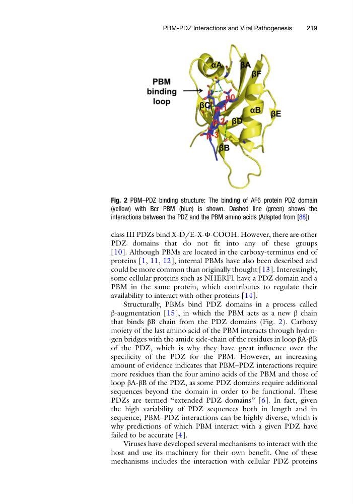
Structurally, PBMs bind PDZ domains in a process called
β-augmentation [15], in which the PBM acts as a new β chain
that binds βB chain from the PDZ domains (Fig. 2). Carboxy
moiety of the last amino acid of the PBM interacts through hydrogen bridges with the amide side-chain of the residues in loop βA-βB
of the PDZ, which is why they have great influence over the
specificity of the PDZ for the PBM. However, an increasing
amount of evidence indicates that PBM–PDZ interactions require
more residues than the four amino acids of the PBM and those of
loop βA-βB of the PDZ, as some PDZ domains require additional
sequences beyond the domain in order to be functional. These
PDZs are termed “extended PDZ domains” [6]. In fact, given
the high variability of PDZ sequences both in length and in
sequence, PBM–PDZ interactions can be highly diverse, which is
why predictions of which PBM interact with a given PDZ have
failed to be accurate [4].
Viruses have developed several mechanisms to interact with the
host and use its machinery for their own benefit. One of these
mechanisms includes the interaction with cellular PDZ proteins
Fig. 2 PBM–PDZ binding structure: The binding of AF6 protein PDZ domain
(yellow) with Bcr PBM (blue) is shown. Dashed line (green) shows the
interactions between the PDZ and the PBM amino acids (Adapted from [88])
PBM-PDZ Interactions and Viral Pathogenesis
219
PDZ domains that do not fit into any of these groups
[10]. Although PBMs are located in the carboxy-terminus end of
proteins [1, 11, 12], internal PBMs have also been described and
could be more common than originally thought [13]. Interestingly,
some cellular proteins such as NHERF1 have a PDZ domain and a
PBM in the same protein, which contributes to regulate their
availability to interact with other proteins [14].
Structurally, PBMs bind PDZ domains in a process called
β-augmentation [15], in which the PBM acts as a new β chain
that binds βB chain from the PDZ domains (Fig. 2). Carboxy
moiety of the last amino acid of the PBM interacts through hydrogen bridges with the amide side-chain of the residues in loop βA-βB
of the PDZ, which is why they have great influence over the
specificity of the PDZ for the PBM. However, an increasing
amount of evidence indicates that PBM–PDZ interactions require
more residues than the four amino acids of the PBM and those of
loop βA-βB of the PDZ, as some PDZ domains require additional
sequences beyond the domain in order to be functional. These
PDZs are termed “extended PDZ domains” [6]. In fact, given
the high variability of PDZ sequences both in length and in
sequence, PBM–PDZ interactions can be highly diverse, which is
why predictions of which PBM interact with a given PDZ have
failed to be accurate [4].
Viruses have developed several mechanisms to interact with the
host and use its machinery for their own benefit. One of these
mechanisms includes the interaction with cellular PDZ proteins
Fig. 2 PBM–PDZ binding structure: The binding of AF6 protein PDZ domain
(yellow) with Bcr PBM (blue) is shown. Dashed line (green) shows the
interactions between the PDZ and the PBM amino acids (Adapted from [88])
PBM-PDZ Interactions and Viral Pathogenesis
219